IMAGE

Fig. 4

ID
ZDB-IMAGE-240531-147
Source
Figures for Inoue et al., 2024
Image
Figure Caption

Fig. 4

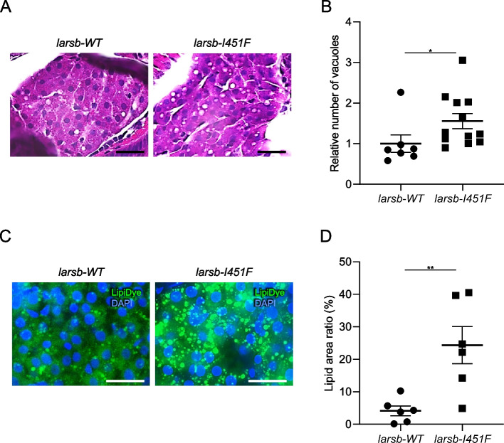
Histopathology and lipids staining of the liver in larsb-knockin larvae. A Hematoxylin and eosin staining of the liver in larsb-knockin (larsb-I451F) larvae at 5 dpf. Scale bar: 20 μm. B Quantification of the vesicle number in larsb-WT (n = 7 sections) and larsb-I451F (n = 12 sections) liver at 5 dpf. Error bars indicate SEM. *P < 0.05. C Lipids staining of the liver in larsb-I451F larvae at 5 dpf. Scale bar: 20 μm. D Quantification of the lipid area in larsb-WT (n = 6 sections) and larsb-I451F (n = 6 sections) larvae liver at 5 dpf. Error bars indicate SEM. **P < 0.01. Dpf: days post fertilization

Acknowledgments
This image is the copyrighted work of the attributed author or publisher, and ZFIN has permission only to display this image to its users. Additional permissions should be obtained from the applicable author or publisher of the image. Full text @ Orphanet J Rare Dis