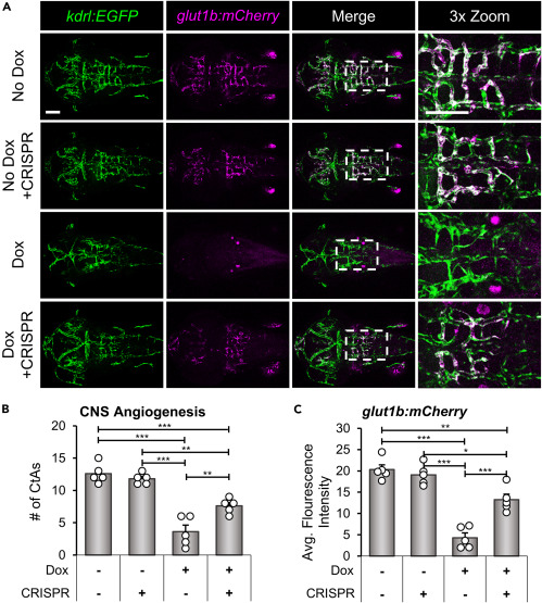
Fig. 5 il1r1 crispants rescue glut1b:mCherry expression in brain endothelial cells during CNS angiogenesis (A) Representative confocal microscopy images showing rescue of glut1b:mCherry expression in il1r1 crispants. CNS/Il-1β, kdrl:EGFP, glut1b:mCherry embryos were injected with CRISPR/Cas9 RNP complexes (cr1 and cr2) at the one-cell stage. Control embryos and il1r1 crispants were either untreated (No Dox) or treated (10.0 μg/mL Dox) at 6 hpf and then imaged at 52 hpf (dorsal view; anterior left). Scale bars are 100 μm. (B and C) Quantification of the number of CtAs (B) and average glut1b:mCherry fluorescence intensity (C) in the hindbrain vasculature of control (CRISPR −) and il1r1 crispants (CRISPR +) either untreated (Dox −) or treated with 10.0 μg/mL Dox (Dox +) (n = 5 for each condition). Error bars in B and C represent means ± SEM (∗p < 0.05; ∗∗p < 0.01; ∗∗∗p < 0.001; no label = not significant).
Image
Figure Caption
Acknowledgments
This image is the copyrighted work of the attributed author or publisher, and
ZFIN has permission only to display this image to its users.
Additional permissions should be obtained from the applicable author or publisher of the image.
Full text @ iScience

