IMAGE

Figure 3

ID
ZDB-IMAGE-240527-11
Genes
Antibodies
Source
Figures for Donizetti et al., 2024
Image
Figure Caption

Figure 3

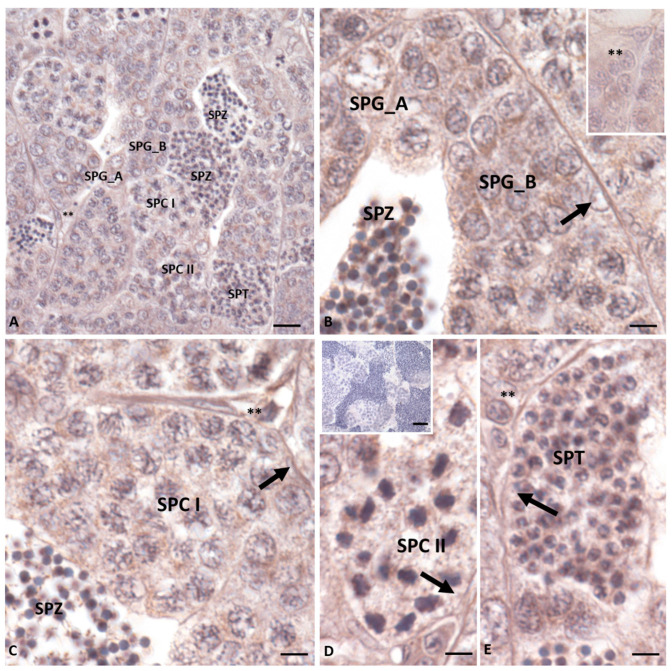
Immunohistochemistry for Insl3 in Danio rerio testis. The immunolocalization signal appears as brown areas. (AE): Antibody positivity is evident in Spermatogonia A (SPG_A), Spermatogonia B (SPG_B), Spermatocytes I (SPC I), Spermatocytes II (SPC II), and Spermatids (SPT) and Spermatozoa (SPZ). The low signal is evident in the LC (asterisks) and no positivity is found in the Sertoli cells (arrow). The negative control sections ((D) inset) show no signal. The scale bars correspond to 50 µm in the figure (D) insert, 20 µm in figure (A), and 10 µm in figures (BE).

Figure Data
Acknowledgments
This image is the copyrighted work of the attributed author or publisher, and ZFIN has permission only to display this image to its users. Additional permissions should be obtained from the applicable author or publisher of the image. Full text @ Int. J. Mol. Sci.