IMAGE

Fig. 2

ID
ZDB-IMAGE-240517-78
Source
Figures for Suppermpool et al., 2024
Image
Figure Caption

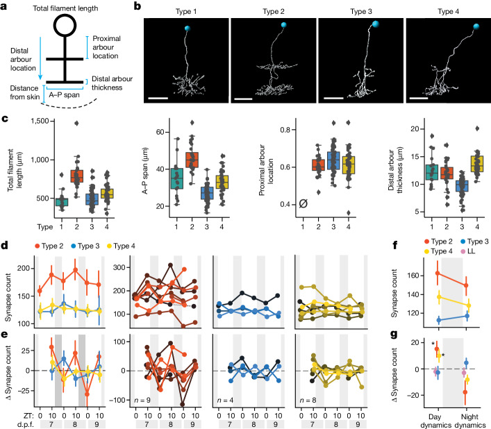
Fig. 2 Subtype-specific synapse changes in FoxP2.A tectal neurons over 3 days.

a, The morphological parameters used to characterize FoxP2.A tectal neurons. A–P, anterior–posterior. b, Examples of each morphological subtype, chosen from n = 17 (type 1), n = 28 (type 2), n = 61 (type 3) and n = 42 (type 4) neurons collected over 26 independent experiments. The blue circles label nuclei. c, Example of the parameters used to distinguish the four subtypes. For the box plots, the centre lines show the median, the box limits show the interquartile range and the whiskers represent the distribution for each parameter. The slashed zero indicates that the feature is absent. See also Extended Data Fig. 5. dg, Synapse counts across multiple LD cycles for FoxP2.A tectal neurons of different subtypes. d,e, Average (68% CI) synapse counts (d) and average (68% CI) synapse number change (e) of subtypes (column 1) and for each neuron (columns 2–4), collected over 8 independent experiments. f,g, Average (68% CI) synapse counts (f) and net change (g), averaged across all days and nights for each subtype and larvae, including additional neurons tracked over a single day (Extended Data Fig. 5). Tectal subtype influences synapse changes (mixed ANOVA, interaction P = 0.012, subtype × time). Type 2 (n = 16) and type 4 (n = 15) neurons gain more synapses during the day under LD conditions compared with under LL clock-break conditions (P = 0.018, g = 0.952; P = 0.021, g = 0.812, respectively). At night, both type 2 and type 4 neurons lose synapses relative to type 3 (type 2 versus type 3, P = 0.038; g = −0.714; type 4 versus type 3, P = 0.038, g = −0.781, post hoc Benjamini–Hochberg correction, one-tailed). For b, scale bars, 10 μm.

Source Data

Acknowledgments
This image is the copyrighted work of the attributed author or publisher, and ZFIN has permission only to display this image to its users. Additional permissions should be obtained from the applicable author or publisher of the image. Full text @ Nature