
|
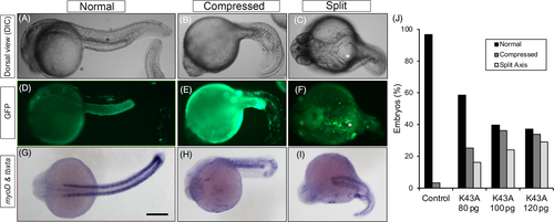
Fig. 6 Ectopic expression of dominant-negative CaMKII induces convergent extension (CE) defects. Embryos were injected with GFP-tagged dominant-negative (K43A) CaMKII cDNA and observed at 24hpf in differential interference contrast light (A–C) and fluorescent (D–F) optics. Embryos with normal, compressed, and split morphologies are shown and were also fixed and probed for tbxta and myoD expression (G–I). Individual fluorescent cells can be seen between the split axes and are marked by an asterisk (C, F). Bar chart represents the accumulated categorization of 58–93 embryos for each level of cDNA injected which spanned seven separate experiments.