
|
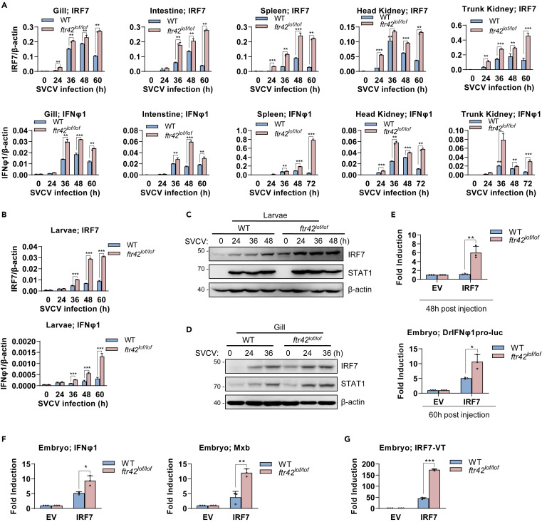
Fig. 7 FTR42 facilitates IRF7 mRNA decay to downregulate IFN response in vivo (A and B) FTR42-dificient zebrafish showed enhanced mRNA expression of irf7 and ifnφ1 over WT zebrafish upon SVCV infection. Zebrafish adults (90 dpf, A) and zebrafish larvae (4 dpf, B) were challenged with SVCV. At the indicated time points, tissues or larvae were sampled for RT-qPCR analyses of cellular ifnφ1 and irf7. (C and D) FTR42-dificient zebrafish showed a higher level of IRF7 protein than WT zebrafish upon SVCV infection. Zebrafish larvae (C) and adults (D) were infected with SVCV. At the indicated time points, larvae or gill were sampled for western blotting analyses of endogenous IRF7 protein. (E) IRF7 stimulated IFN promoter activation more significantly in ftr42lof/lof embryos than in WT embryos. One-cell-stage embryos from WT and FTR42-dificient zebrafish were microinjected with IRF7 (50 pg), DrIFNφ1pro-luc (50 pg) and pRL-TK (2.5 pg), in a volume of 1 nL per embryo. At the indicated time points, the embryos were collected for luciferase assays. (F) IRF7 induced transcriptional expression of ifnφ1 and mxb more significantly in ftr42lof/lof embryos than in WT embryos. One-cell-stage embryos were microinjected with IRF7 (60 pg), in a volume of 1 nL per embryo. 48 h later, ifnφ1 and mxb mRNA was detected by RT-qPCR. (G) FTR42-dificient zebrafish expressed a higher level of IRF7 mRNA than WT zebrafish after microinjection. The same samples in (F) were used to detect irf7 mRNA from the microinjected IRF7 plasmid by RT-qPCR. Data were shown as mean ± SD (N = 3). p values were calculated using Student’s t test. ∗p < 0.05, ∗∗p < 0.01, ∗∗∗p < 0.001.