
|
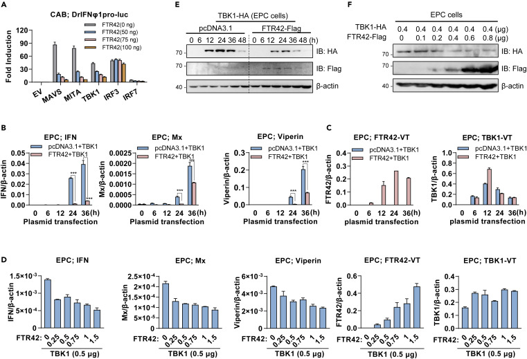
Fig. 3 FTR42 impairs IFN response by attenuating TBK1 protein level in vitro (A) Overexpression of FTR42 inhibited IFN promoter activation by MAVS, MITA, TBK1 and IRF7 but not by IRF3. CAB cells seeded in 48-well plates overnight were transfected with DrIFNφ1pro-luc, each of signaling molecules (MAVS, MITA, TBK1, IRF3 and IRF7) or empty vector as control (100 ng each), together with FTR42 at increasing amounts. 10 ng pRL-TK was included as internal control. 30 h later, cells were harvested for luciferase assays. (B–D) FTR42 downregulated TBK1-trigged IFN response. EPC cells seeded in 6-well plates were transfected with the indicated plasmids as in (A), and were then collected at the indicated time points (B and C) or at 30 h post transfection (D), for RT-qPCR analyses of mRNA expression of cellular ifn and ISGs (B and D), and mRNA expression from the transfected FTR42-vt and TBK1-vt (C and D). (E and F) FTR42 attenuated TBK1 protein levels in vitro. EPC cells seeded in 3.5 cm2 dishes overnight were transfected with TBK1, FTR42 or pcDNA3.1 as control (1 μg each), and at different time points, the cells were harvested for western blotting analyses (E). Or the cells were transfected for 30 h with TBK1 and FTR42 at the indicated doses, followed by western blotting (F). Data were shown as mean ± SD (N = 3). p values were calculated using Student’s t test. ∗∗∗p < 0.001.