
|
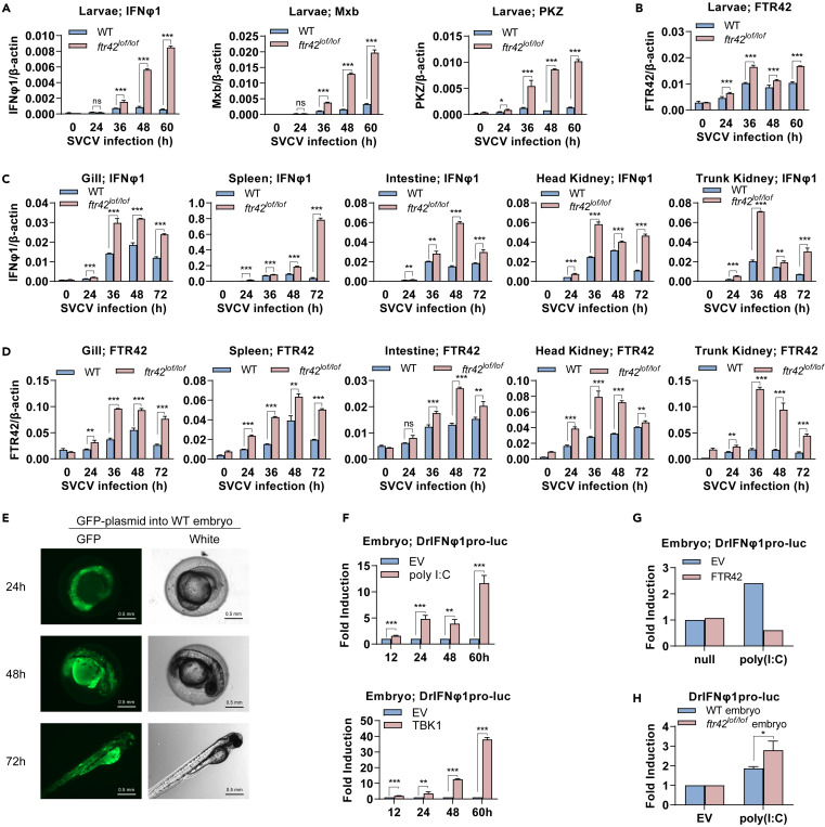
Fig. 2 . FTR42-deficient zebrafish show an enhanced IFN immunity (A and B) FTR42-deficient zebrafish larvae showed an enhanced IFN immunity. WT and ftr42lof/lof larvae (4 dpf) were immersed with SVCV (5×10⁶ TCID50/mL) for indicated time points, followed by RT-qPCR analyses of ifnφ1, mxb and pkz (A) and ftr42 (B). (C and D) FTR42-deficient zebrafish adults showed an enhanced IFN immunity. WT and ftr42loflof adults (90 dpf) were i.p. injected with SVCV (1×10⁸ TCID50/mL) for indicated time points, followed by RT-qPCR analyses of ifnφ1 (C) and ftr42 (D) in various tissues. (E) GFP was continuously expressed in zebrafish embryos by microinjection. A GFP plasmid (60 pg/nL) was microinjected into one-cell-stage zebrafish embryos (1 nL per embryo), followed by fluorescence microscopy at indicated time points. Scale bar: 0.5 mm. (F) IFNφ1 promoter was activated in zebrafish embryos by poly(I:C) or TBK1. One-cell-stage embryos were microinjected with DrIFNφ1pro-luc (50 pg/nL) and pRL-TK (2.5 pg/nL), together with poly(I:C) (50 pg/nL) or TBK1 (50 pg/nL), in a total volume of 1 nL per embryo. At the indicated time points, embryos were collected for luciferase assays. (G and H) IFNφ1 promoter activation was inhibited in zebrafish embryos by FTR42. One-cell-stage WT embryos were microinjected as in (F) with DrIFNφ1pro-luc, poly(I:C) and FTR42 (50 pg/nL), together with pRL-TK (2.5 pg/nL) (G). Or WT and ftr42lof/lof embryos were injected with DrIFNφ1pro-luc, poly(I:C) and pRL-TK, respectively (H). 60 h later, luciferase assays were performed. Data were shown as mean ± SD (N = 3). p values were calculated using Student’s t test. ∗p < 0.05, ∗∗p < 0.01, ∗∗∗p < 0.001.