IMAGE

Fig 6

ID
ZDB-IMAGE-240511-6
Source
Figures for Ahuja et al., 2024
Image
Figure Caption

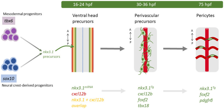
Fig 6 nkx3.1 is essential in pericyte precursors.

Model of nkx3.1 in pericyte differentiation. nkx3.1 is expressed in cells of both mesodermal and neural crest origin as determined by lineage analysis. All embryo schematics show a dorsal view of the hindbrain. At 16 and 24 hpf, nkx3.1 and cxcl12b coexpressing precursors are located ventrolaterally. At 36 hpf, nkx3.1, cxcl12b, tbx18, and foxf2 coexpressing precursors surround the developing basilar artery (BA). By 75 hpf, pdgfrβ-expressing pericytes derived from nkx3.1 precursors have migrated to the central arteries of the brain. Key genetic markers are indicated.

Acknowledgments
This image is the copyrighted work of the attributed author or publisher, and ZFIN has permission only to display this image to its users. Additional permissions should be obtained from the applicable author or publisher of the image. Full text @ PLoS Biol.