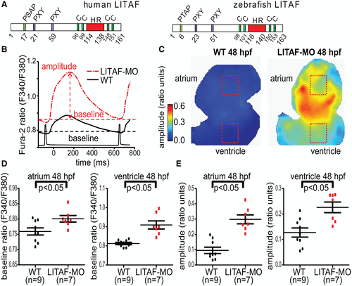
Fig. 1 Genetic knockdown of LITAF (lipopolysaccharide-induced tumor necrosis factor) increases calcium transients in the heart of zebrafish embryos. Zebrafish hearts from 48 hpf (hours post-fertilization) wild-type (WT) and LITAF morphants (MO) were stained with Fura-2 AM (Fura-2-acetoxymethyl ester) to measure Ca2+ transients. A, Structure of human and zebrafish LITAF with conserved PXY and P(S/T)AP motifs and the hydrophobic region (HR) required for membrane embedding.49 Also indicated are 2 conserved cysteine pairs for coordination of a zinc atom likely required for proper protein folding.49 B, Averaged ventricular Ca2+ transients (amplitudes and baselines) from regions of interest (ROIs) as indicated in the color maps. C, Color maps of Ca2+ transient amplitudes from WT and LITAF morphant hearts. Color code depicts Ca2+ transient amplitudes in fluorescence ratio units (F340/F380). Squares indicate ROIs for measurements averaged in B. D, Mean baseline ratios. E, Mean Ca2+ transient amplitudes. Error bars depict SEM. One-way ANOVA, *P<0.05 for comparisons with WT (n=9 wild-type zebrafish embryos; n=7 LITAF morphants).
Image
Figure Caption
Acknowledgments
This image is the copyrighted work of the attributed author or publisher, and
ZFIN has permission only to display this image to its users.
Additional permissions should be obtained from the applicable author or publisher of the image.
Full text @ Circ Genom Precis Med

