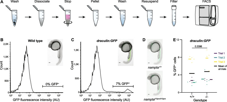
nampt-a+/+ and nampt-at10pm/t10pm embryos have comparable draculin:GFP-positive cell populations. A To quantify lateral plate mesoderm cells, draculin:GFP zebrafish embryos were homogenized, and GFP+ cells were counted using fluorescence activated cell sorting (FACS). B Wild-type embryos not expressing GFP were used as controls to establish a cut-off for GFP− cells. C Example of FACS read-out of GFP+ cells (green, inset) in draculin:GFP embryos that are detected above the baseline cut-off set in (B). D Images of nampt-a+/+ and nampt-at10pm/t10pm embryos in the draculin:GFP background before homogenization and quantification by FACS. E Quantification of GFP+ cells in nampt-a+/+ (+ / +) and nampt-at10pm/t10pm (-/-) embryos. Each point represents one biological sample of ten pooled embryos. Each colored line represents the average from separate experimental trial days, and the black bar shows the mean of all trials. For statistical analysis, the Mann–Whitney test was applied to determine the p-value. Scale bars in B-D represent 100 µm
This image is the copyrighted work of the attributed author or publisher, and
ZFIN has permission only to display this image to its users.
Additional permissions should be obtained from the applicable author or publisher of the image.
Full text @ Hereditas
Your Input Welcome
Thank you for submitting comments. Your input has been emailed to ZFIN curators who may contact you if
additional information is required.
Oops. Something went wrong. Please try again later.