
Fig. 5
- ID
- ZDB-IMAGE-240319-28
- Publication
- Megerson et al., 2023 - Kremen1 regulates the regenerative capacity of support cells and mechanosensory hair cells in the zebrafish lateral line
- All Figures
- Figures for Megerson et al., 2023
|
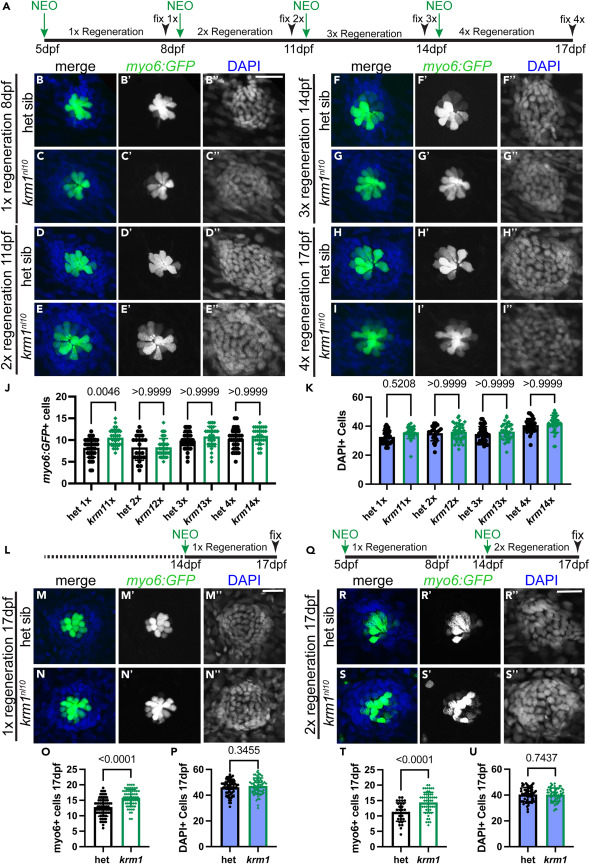
Fig. 5 Repeated regeneration eliminates supernumerary hair cells in krm1nl10 larvae (A) Timeline of NEO exposure and repeated regeneration. (B–I″) Confocal projections of NMs expressing Tg(myosin6b:GFP)w186 in hair cells (green) and DAPI-labeling in nuclei following repeated exposures to NEO in heterozygous sibling and krm1nl10 mutant NMs at 8dpf following 1 round of NEO exposure and regeneration, at 11dpf following 2 rounds of NEO exposure and regeneration, at 14dpf following 3 rounds of NEO exposure and regeneration, and at 17dpf following 4 rounds of NEO exposure and regeneration. (J–K) Quantification of regenerated Tg(myosin6b:GFP)w186 hair cells or DAPI-labeled nuclei in heterozygous sibling and krm1nl10 mutant NMs at 8dpf, 11dpf, 14dpf, and 17dpf. 8dpf heterozygous sibling n = 38 NM (8 fish) and krm1nl10 mutant n = 30 NM (10 fish), 11dpf heterozygous sibling n = 23 (9 fish) and krm1nl10 n = 39 NM (10 fish), 14dpf heterozygous sibling n = 37 NM (9 fish) and krm1nl10 n = 31 NM (9 fish), and 17dpf heterozygous sibling n = 34 NM (10 fish) and krm1nl10 n = 31 NM (9 fish). (L) Timeline of a single round NEO-exposure and regeneration between 14 and 17dpf. (M–N″) Heterozygous sibling and krm1nl10 mutant NMs at 17dpf following 1 round of NEO exposure and regeneration. (O–P) Quantification of regenerated Tg(myosin6b:GFP)w186 hair cells and DAPI-positive cells at 17dpf. Heterozygous siblings n = 68 NM (12 fish) and krm1nl10 n = 62 NM (12 fish). (Q) Timeline of 2 rounds of NEO exposure (at 5dpf and 14dpf) and regeneration until 17dpf. (R–S″) Heterozygous sibling and krm1nl10 mutant NMs at 17dpf following 2x rounds of NEO exposure and regeneration. (T–U) Quantification of regenerated Tg(myosin6b:GFP)w186 hair cells and DAPI-positive cells at 17dpf. Heterozygous siblings n = 33 NM (8 fish) and krm1nl10 n = 58 NM (9 fish). All data presented as mean ± SD, Kruskal-Wallis test, Dunn’s multiple comparisons test. Scale bar = 20 μm.