
Fig. 5
- ID
- ZDB-IMAGE-240314-12
- Publication
- Chiang et al., 2023 - The Role of MAPRE2 and Microtubules in Maintaining Normal Ventricular Conduction
- All Figures
- Figures for Chiang et al., 2023
|
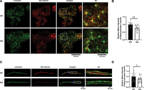
Fig. 5 mapre2 loss of function leads to decreased detyrosinated tubulin. A, Representative immunostaining of hearts from wild-type (WT; n=8) and homozygous knockout (KO; n=16) larvae showing a decrease in ventricular detyrosinated tubulin (Glu-tubulin) relative to total α-tubulin. B, Quantification of ventricular Glu-tubulin signal using α-tubulin signal as a mask (unpaired t test, P=0.0072). C, Representative immunostaining of ventricular myocytes isolated from adult WT (21 cells from 2 fish) and homozygous KO fish (20 cells from 3 fish) also showing a decrease in ventricular detyrosinated tubulin (Glu-tubulin) relative to total α-tubulin. D, Quantification of ventricular Glu-tubulin signal using α-tubulin signal as a mask (unpaired t test, P=0.0232). Representative images were chosen based on closeness to group mean and image quality.