IMAGE

Figure 2

ID
ZDB-IMAGE-240305-22
Source
Figures for Lee et al., 2024
Image
Figure Caption

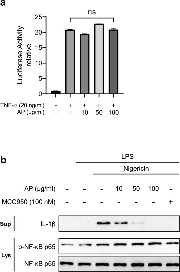
Figure 2

AP does not affect the NF-κB signaling pathway. (a) 293 T cells were transfected with the pGL4.32 luciferase reporter vector and treated with TNF-α (20 ng/mL) for 5 h with or without AP. Luciferase activity was analyzed using the Promega Bright-Glo™ Luciferase Assay System. (b) LPS-primed J774A.1 cells were treated with AP for 2 h and activated for 30 min with nigericin (10 μM). IL-1β in the supernatants (Sup), phospho-NF-κB and NF-κB in the soluble lysates (Lys) were analyzed using a Western blot.

Acknowledgments
This image is the copyrighted work of the attributed author or publisher, and ZFIN has permission only to display this image to its users. Additional permissions should be obtained from the applicable author or publisher of the image. Full text @ Sci. Rep.