IMAGE

Fig. 4.

ID
ZDB-IMAGE-240305-12
Source
Figures for Iyer et al., 2024
Image
Figure Caption

Fig. 4.

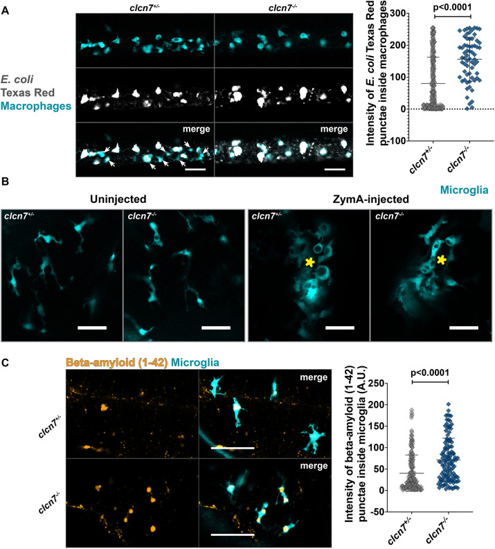
Exogenous debris accumulates inside microglia and macrophages of clcn7 mutants. (A) E. coli Texas Red particles injected in the ducts of Cuvier accumulate inside peripheral macrophages. The intensity of Texas Red particles inside the macrophages is greater in clcn7 mutants (n=8, mean=156.2, s.d.=68.8, s.e.m.=8.5) than in clcn7 heterozygotes (n=27, mean=80.2, s.d.=82.3, s.e.m.=5.1). Arrows indicate macrophages containing little or no E. coli in control animals. Scale bars: 50 µm. (B) Zymosan A injected into the midbrain of zebrafish larvae to observe microglia response. Microglia in clcn7 mutants and heterozygotes respond similarly to inflammatory stress. Yellow asterisks mark the site of injection. Images are representative of two independent experiments. Scale bars: 20 µm. (C) β-amyloid (1–42) HiLyte Fluor-555 particles injected into the midbrain of zebrafish larvae accumulate inside the microglia of clcn7 mutants (n=16, mean=71.7, s.d.=50.3, s.e.m.=4.6) to a significantly greater extent than in clcn7 heterozygotes (n=26, mean=40.5, s.d.=42.5, s.e.m.=3.0). Scale bars: 50 µm. Two-tailed unpaired t-test with Welch's correction was performed to calculate the significance in all graphs; graphs show mean±s.d.

Figure Data
Acknowledgments
This image is the copyrighted work of the attributed author or publisher, and ZFIN has permission only to display this image to its users. Additional permissions should be obtained from the applicable author or publisher of the image. Full text @ J. Cell Sci.