IMAGE

Fig. 3

ID
ZDB-IMAGE-240304-9
Source
Figures for Stegmann et al., 2024
Image
Figure Caption

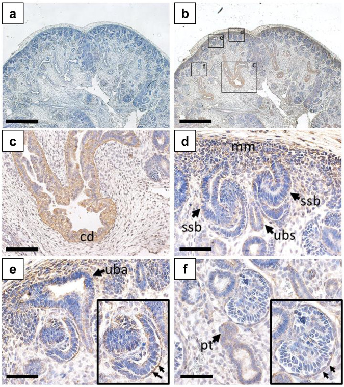
Fig. 3 CELSR3 immunostaining in the human embryonic metanephric kidney at ten weeks gestation.

All frames depict a ten-week gestation kidney with nuclei counterstained (blue) with hematoxylin. a Low power view of midsagittal section with primary antibody omitted. The nephrogenic cortex is uppermost and the medulla is in the low part of the image. Note the absence of brown color. b Adjacent section to that depicted in a. but immuno-stained for CELSR3. Note the positive signal (brown) in diverse structures. Boxed areas are detailed in c-f. c CELSR3 was detected in branching medullary collecting ducts (cd). d The nephrogenic cortex contains immature structures. CELSR3 was detected in the ureteric bud branch stalk (ubs) which is flanked by nephron precursors called S-shape bodies (ssb). The metanephric mesenchyme (mm) stained weakly for CELSR3. e Another view of the nephrogenic cortex showing the ureteric bud branch ampullary tip (uba). These epithelia were weakly positive for CELSR3. Lower in the same image is an immature glomerulus with prominent CELSR3 immunostaining in the Bowman capsule, or parietal epithelia (arrows in the boxed enlargement). f The Bowman capsule of a more mature glomerulus has downregulated CELSR3 (arrows in boxed enlargement), and there is weak immunostaining in a nearby proximal tubule (pt). Bars are 2 mm in frames a and b, and 200 µm in frames cf.

Acknowledgments
This image is the copyrighted work of the attributed author or publisher, and ZFIN has permission only to display this image to its users. Additional permissions should be obtained from the applicable author or publisher of the image. Full text @ NPJ Genom Med