
|
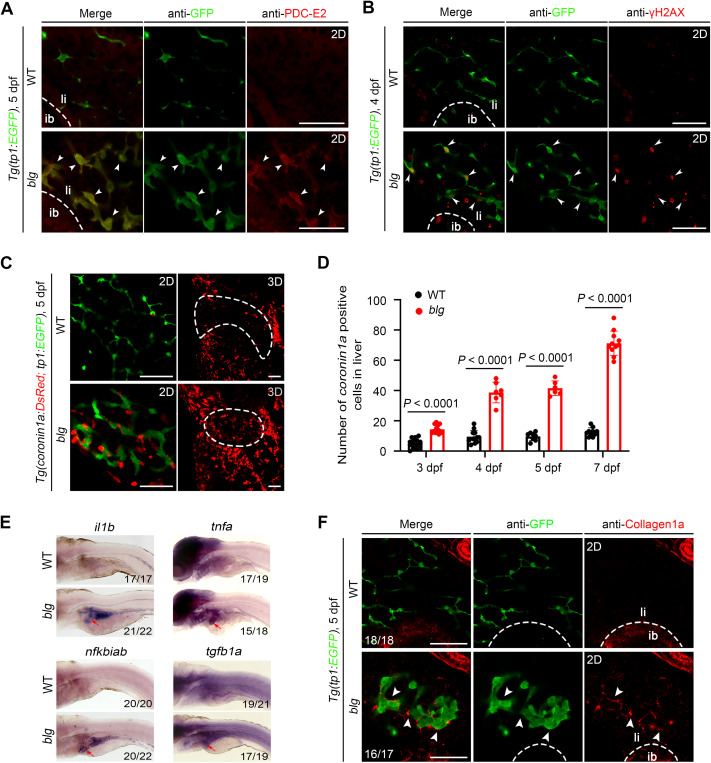
Fig. 3 The blg/ppp1r21 mutant develops analogous pathological features of PBC. A: Immunofluorescence staining of PDC-E2 in the WT and blg mutant under Tg(tp1:EGFP) background at 5 dpf. Arrowheads indicate increased PDC-E2 in blg cholangiocytes. B: Immunofluorescence staining showed elevated levels of γH2AX in the blg mutant compared to the WT at 4 dpf. Arrowheads indicate γH2AX positively overlap with blg cholangiocytes. C: Confocal 3D projection and 2D single-optical section images of WT or blg mutant at 5 dpf under Tg(tp1:EGFP, coronin1a:DsRed) background. coronin1a positive cells (red) were gathered around the bile duct (green) in the blg mutant. The dotted outlines indicate the liver area. D: Statistical diagram of the number of the DsRed positive cells in the WT and blg mutant (per group n ≥ 6) from 3–7 dpf under Tg(coronin1a:DsRed) background. Data are expressed as mean ± SEM, Student's t-test. E: Whole-mount in situ hybridization showed that expressions of il1b, tnfa, nfkbiab, and tgfb1a were up-regulated in the liver (arrows) of blg mutant compared to the WT larvae at 5 dpf. Lateral views, anterior left. F: Immunofluorescence staining of Collagen1a in the WT and blg mutant at 5 dpf under Tg(tp1:EGFP) background. Arrowheads indicate increased Collagen1a around the blg cholangiocytes. WT, wild-type; ib, intestinal bulb; li, liver. Scale bars, 50 μm (A–C, F). n = number of embryos with indicated phenotype/total analyzed in each class.
Reprinted from Journal of genetics and genomics = Yi chuan xue bao, 50(12), Wu, C., Zhang, W., Luo, Y., Cheng, C., Wang, X., Jiang, Y., Li, S., Luo, L., Yang, Y., Zebrafish ppp1r21 mutant as a model for the study of primary biliary cholangitis, 1004-1013, Copyright (2023) with permission from Elsevier. Full text @ J. Genet. Genomics