
|
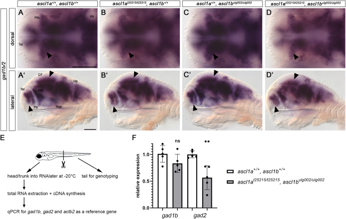
Fig. 5 Ascl1 is required for proper GABAergic neurogenesis. (A-D′) Whole-mount in situ hybridization for gad1b/2 on 96 hpf embryos. Dorsal (A–D) and lateral views (A′-D′) of wildtype, single and double mutant ascl1a and ascl1b embryos. Anterior is to the left. Scale bars: 100 μm. Absence of Ascl1 activity leads to a reduction of GABAergic neurons visualized by gad1b/2 expression in the prethalamus, telencephalon and optic tectum (arrowheads, A-D′). In contrast, the expression of gad1b/2 appears normal in the hindbrain in all analyzed genotypes. For numbers of analyzed embryos see Supplemental Table S2. Abbreviations: HB, hindbrain; Hyp, hypothalamus; OT, optic tectum; Po, preoptic region; Pth, prethalamus; Tel, telencephalon. (E,F) qPCR for gad1b and gad2 expression in 96 hpf embryos. (E) 96 hpf embryos were tail-clipped and processed as depicted in the schematic. (F) The expression of gad1b and gad2 is reduced in double mutant embryos compared to wildtype, but only significantly in case of gad2. qPCRs were performed with five biological replicates per genotype, and measurements with three technical replicates, dots show biological replicates. (p = 0.0024; unpaired t-test on ΔCq values). Error bars - standard error of the mean.
Reprinted from Developmental Biology, 505, Altbürger, C., Rath, M., Wehrle, J., Driever, W., The proneural factors Ascl1a and Ascl1b contribute to the terminal differentiation of dopaminergic GABAergic dual transmitter neurons in zebrafish, 587458-74, Copyright (2023) with permission from Elsevier. Full text @ Dev. Biol.