
|
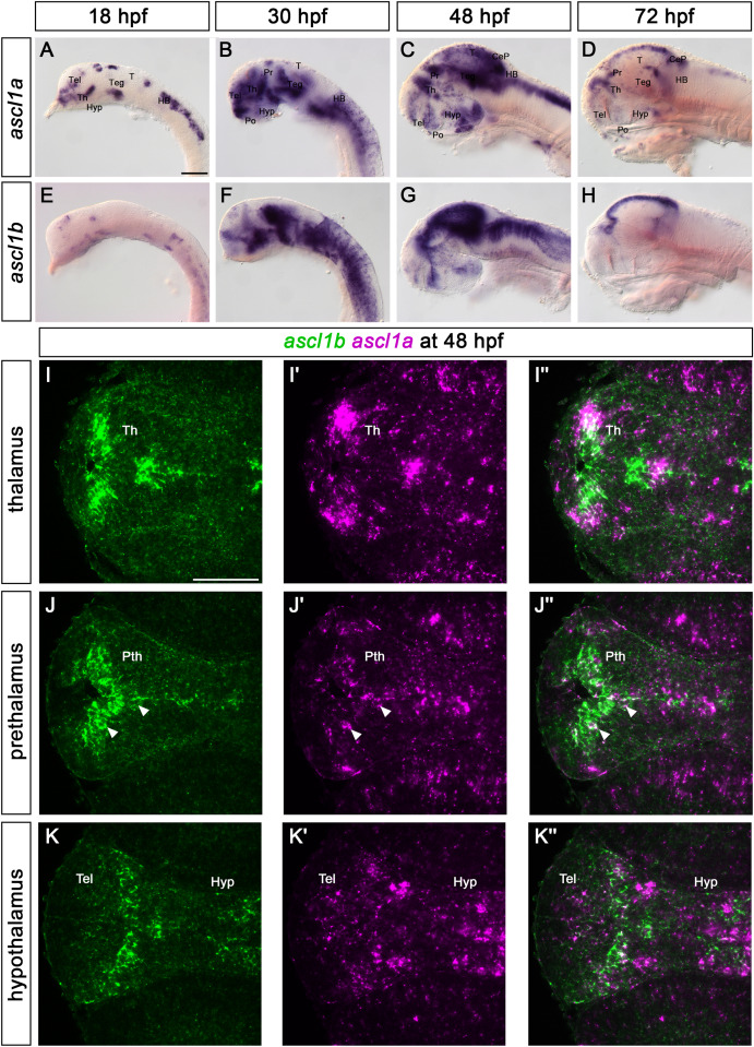
Fig. 1 The expression of ascl1a and ascl1b throughout development. (A–H) Whole-mount in situ hybridizations for ascl1a (A–D) and ascl1b (E–H). Lateral views of wildtype embryos at the indicated stages. Anterior is to the left. (I–K″) Fluorescent whole mount in situ hybridization of ascl1a and ascl1b at 48 hpf. Dorsal views of single confocal planes of wildtype embryos. Anterior is to the left. The expression of ascl1a and ascl1b is mainly exclusive in the thalamus (I–I″) and hypothalamus (K–K″). In the prethalamus (J-J″) a few cells lining or being close to the ventricle express both paralogs (white arrowheads). The confocal stack is provided as Supplemental Video S1. Abbreviations: CeP, cerebellar plate; HB, hindbrain; Hyp, hypothalamus; Po, preoptic region; Pr, pretectum; Pth, prethalamus; T, tectum; Teg, tegmentum; Tel, telencephalon; Th, thalamus. Scale bars: 100 μm.
Reprinted from Developmental Biology, 505, Altbürger, C., Rath, M., Wehrle, J., Driever, W., The proneural factors Ascl1a and Ascl1b contribute to the terminal differentiation of dopaminergic GABAergic dual transmitter neurons in zebrafish, 587458-74, Copyright (2023) with permission from Elsevier. Full text @ Dev. Biol.