IMAGE

Fig. 1

ID
ZDB-IMAGE-240229-171
Source
Figures for Veloso et al., 2024
Image
Figure Caption

Fig. 1

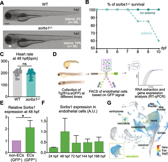
Sorbs1 expression is enriched in the endothelium and its deletion in zebrafish results in cardiac edemas. A Transmitted light images of live wild-type (WT) and sorbs1 mutant (sorbs1−/−) zebrafish embryos at 5 days post-fertilization (dpf). The white arrow indicates an example of edema observed in sorbs1−/− embryo. Proportions of embryos with edemas in each genotype are indicated. Scale bar represents 250 μm. B Quantification of the percentage of survival for sorbs1−/− embryos presenting or not edemas from 4 to 10 dpf (n = 23 and n = 47, respectively). C Quantification of the heart rate in WT and sorbs1−/− embryos at 48 hpf (n = 45 and n = 49, respectively). D Illustration of the procedure used to sort endothelial cells by FACS (fluorescence-activated cell sorting) from Tg(fli1a:eGFP)y1 zebrafish embryos, at different stages of development, followed by RNA extraction and reverse transcription-qPCR (RT-qPCR). E, F RT-qPCR analysis of sorbs1 expression relative to Elfa after FACS sorting as described in D, in endothelial cells (ECs, GFP + , green bars) vs non-endothelial cells (non-ECs, GFP-, purple bar) (E) or in endothelial cells at different time points of embryonic development (F) (*P < 0.05, unpaired t-test). G UMAP of sorbs1 expression at single-cell level obtained from the Daniocell database consisting of whole-animal wild-type zebrafish across multiple stages of development (14–120 hpf). Cluster annotation was taken from the Daniocell resource

Acknowledgments
This image is the copyrighted work of the attributed author or publisher, and ZFIN has permission only to display this image to its users. Additional permissions should be obtained from the applicable author or publisher of the image. Full text @ BMC Biol.