
|
Fig 6 Boundary and loading conditions in the FE model.
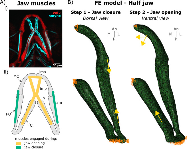
A) Lower jaw muscles. (i) Maximum projection of ventral confocal image stacks expressing Col2a1aBAC:mcherry (red) and smyhc1:EGFP (cyan) of a 4 dpf larva. (ii) Schematic of the muscles engaged during lower jaw opening (yellow) and closure (green) in the ventral plane. am: adductor mandibularis, ih: interhyal, ima: intermandibularis anterior, imp: intermandibularis posterior. B) Half jaw finite element (FE) model of jaw closure and opening with boundary conditions and muscle loads. An: Anterior, CH: ceratohyal, L: Lateral, M: Medial, MC: Meckel’s cartilage, P: Posterior. PQ: palatoquadrate.