
|
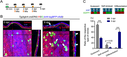
Fig. 6 Induction of basal cell differentiation by non-basal keratinocyte injury. (A) Experimental procedure of basal cell labelling and krt4+ cell injury. Mtz, metronidazole treatment. (B) Confocal images and their optical sections of the EGFP-labelled basal cells in the adult fin before (left panel) and after (6 dpt, right panel) non-basal cell injury. Thin white arrows indicate posterior side (P). Dashed lines indicate basement membrane. After injury of krt4+ cells, the basal cells often became the krt4+ keratinocytes at 6 dpt. Note that such clones did not contain the basal cells, indicating that the basal cells differentiated into non-basal cells (thick white arrows). Scale bar: 20 µm. (C) Quantification of the basal cell reactions in response to non-basal cell injury. The number of clones was counted on confocal images (n=156 clones at 0 dpt, n=276 clones at 6 dpt from four adult fish fins). Respective proliferative patterns are schematically shown above the graph. Error bars denote mean±s.e.m. ***P<0.001 (paired two-tailed Student's t-test). dpt, days post treatment.