IMAGE

Figure 1

ID
ZDB-IMAGE-240215-98
Source
Figures for Raas et al., 2024
Image
Figure Caption

Figure 1

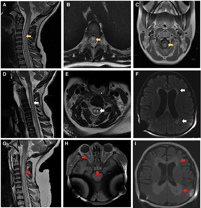
Brain and spinal cord imaging. At initial presentation: (A) MRI cervical spine sagittal T2-weighted image with hyperintense signal involving the central gray matter, dorsal and lateral tracts of the cervical and thoracic spinal cord (yellow arrow). (B) MRI thoracic spine axial T2-weighted image with hyperintense signal involving primarily gray matter (yellow arrow). (C) MRI cervical spine axial post-gadolinium T1-weighted image with contrast enhancement of the dorsal white matter tracts (yellow arrow). Follow-up at 6 months: (D) MRI cervical spine sagittal T2-weighted image with re-demonstration of central gray matter hyperintensity of the cervical and thoracic spinal cord with associated volume loss (white arrow). (E) MRI cervical spine axial T2-weighted image with increased hyperintense signal involving primarily gray matter and volume loss within the gray matter tracts (white arrow). (F) MRI brain axial T2-weighted FLAIR image with diffuse parenchymal volume loss and nearly symmetric bilateral periventricular and deep white matter signal abnormality in the frontal, parietal, and occipital lobes (white arrows). Obtained 2 weeks before death: (G) MRI cervical spine sagittal T2-weighted image with recurrent non-enhancing hyperintense signal involving primarily the dorsal central aspect of the spinal (red arrow). (H) MRI brain axial T1-weighted imaging post-gadolinium with contrast enhancement involving the optic chiasm, prechiasmatic optic nerves, and bilateral optic tracts within the suprasellar region (red arrows). (I) MRI brain axial T2-weighted FLAIR image with progressive bilateral periventricular white matter signal abnormality with central areas of white matter necrosis (red arrows).

Acknowledgments
This image is the copyrighted work of the attributed author or publisher, and ZFIN has permission only to display this image to its users. Additional permissions should be obtained from the applicable author or publisher of the image. Full text @ Front Pediatr