
Fig. 2
- ID
- ZDB-IMAGE-240215-24
- Publication
- Sundaramurthi et al., 2023 - Ergolide mediates anti-cancer effects on metastatic uveal melanoma cells and modulates their cellular and extracellular vesicle proteomes
- All Figures
- Figures for Sundaramurthi et al., 2023
|
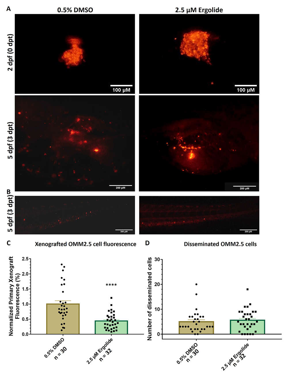
Fig. 2 Anti-cancer effects of ergolide observed in vivo in zebrafish OMM2.5 xenograft models. (A, C) Representative images of zebrafish larvae transplanted with OMM2.5Dil labelled cells at 2 days post fertilization (dpf, top panel); representative images of zebrafish larvae transplanted with OMM2.5Dil labelled cells at 5 dpf, 3 days post treatment (dpt, bottom panel). A 56% (****, p<0.0001%) reduction, on average, in primary xenograft fluorescence observed in the 2.5 μM ergolide (n = 32) treated samples compared to 0.5% DMSO (n = 30) treated samples. (B, D) A significant difference was not detected in the average number of disseminated cells. Statistical analysis performed using Student's T-test.