IMAGE

Figure 4

ID
ZDB-IMAGE-240215-100
Source
Figures for Raas et al., 2024
Image
Figure Caption

Figure 4

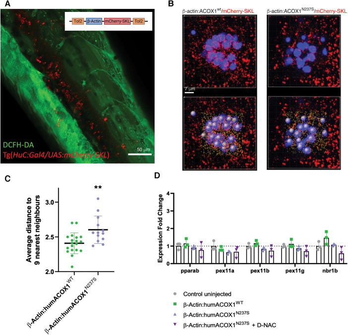
Zebrafish peroxisomal reporter construct and characterization of effects on peroxisomes. (A) Schematic of the mCherry-SKL construct and transient expression in zebrafish. Confocal z-stack, lateral view of the trunk, live 3 dpf larva, green channel, DCFH-DA (dacetyldichlorofluorescein). (B) Live confocal images and paired IMARIS (3D reconstruction) image (below), of mCherry-SKL (red) containing peroxisomes in the hair cells (blue) of larval zebrafish neuromast at 6 dpf in zebrafish overexpressing WT or N237S ACOX1. (C) Size and distance to nearest neighbors in zebrafish overexpressing WT or N237S ACOX1, showing a decreased density of peroxisomes in these cells. (D) qRT-PCR for expression of genes that affect the abundance of peroxisome: pparab, pex11a, pex11b, pex11g, and nrb1b.

Acknowledgments
This image is the copyrighted work of the attributed author or publisher, and ZFIN has permission only to display this image to its users. Additional permissions should be obtained from the applicable author or publisher of the image. Full text @ Front Pediatr