IMAGE

Figure 6

ID
ZDB-IMAGE-240212-24
Source
Figures for Napoli et al., 2024
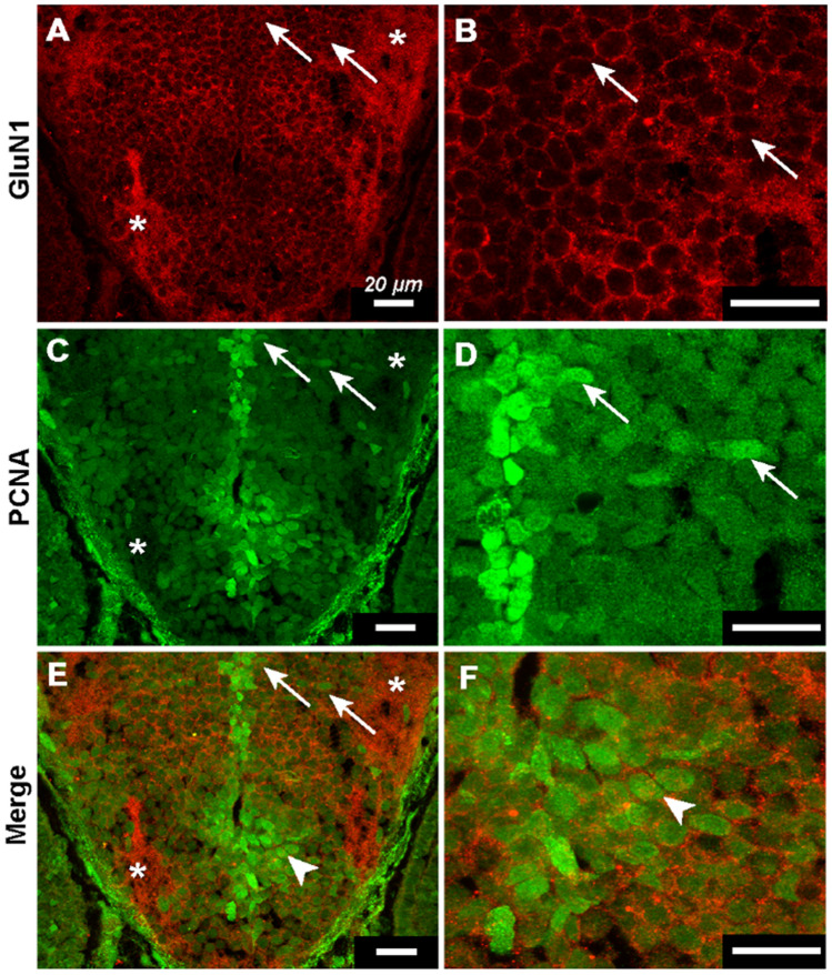
Image
Figure Caption

Figure 6

Transit amplifying cells express GluN1. (AF) Immunohistochemistry against GluN1 and PCNA in 3 dpf wild-type fish. Panels on the right are magnified versions of those on the left. Asterisks indicate example regions of neuropil in (A, C, & E). (A & B) GluN1 channel showing robust plasmelemmal and neuropilar dendritic expression of GluN1. Arrows indicate example cells in abventicular and cell-rich parenchymal locations (A) and illustrate plasmelemmal GluN1 expression (B). (C & D) PCNA channel showing ventricular PCNA+ NSCs and abventricular and cell-rich parenchymal PCNA+ tACs cells. Arrows indicate the same cells as in (A & B) and illustrate that the same cells express both PCNA and GluN1. (E & F) Merged GluN1 and PCNA channels. Arrows indicate same cells as in (A, B, C, & D) arrow heads indicate example PCNA+ tAC with GluN1 puncta.

Acknowledgments
This image is the copyrighted work of the attributed author or publisher, and ZFIN has permission only to display this image to its users. Additional permissions should be obtained from the applicable author or publisher of the image. Full text @ Sci. Rep.