IMAGE

Figure 1

ID
ZDB-IMAGE-240212-17
Source
Figures for Napoli et al., 2024
Image
Figure Caption

Figure 1

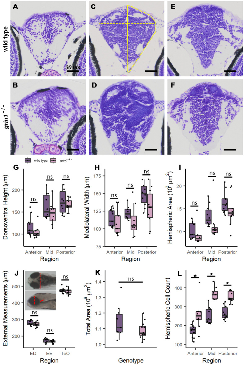
grin1−/− fish do not show gross morphological changes in brain structure. (AF) Nissl stained 5-micron coronal sections of a 5 dpf wild-type (A, C, & E) and a grin1−/− (B, D, & F) fish at the anterior (A & B), mid (C & D) and posterior (E & F) forebrain. Measurement locations indicated in (C): yellow solid line, dorsoventral height measured at midline; dashed line, mediolateral width measured at widest aspect of the brain; and dotted line, hemispheric area. (GI) Box and whisker plots indicating median value, interquartile ranges, and maximum and minimum values, for coronal morphometric measurements of dorsoventral height (G), mediolateral width (H) and hemispheric area (I). In this and all subsequent figures, points represent individual values for each fish. (J & K) External brain size proxy measurements (J) and total area of fish measured in lateral view (K). Inset to (J): dorsal (upper) and lateral (lower) views with measurement locations indicated in red dashed line (optic tectum width, TeO), dotted line (eye diameter, ED), and solid line (eye to ear distance, EE). (l) Total hemispheric cell counts of 5 dpf fish performed on representative sections at anterior, mid, and posterior levels of the forebrain (A–F) in wild-type and grin1−/− fish. Significance (t-test) is indicated (*p < 0.05; ns, not significant). For specific sample sizes and statistical values see Table 2.

Acknowledgments
This image is the copyrighted work of the attributed author or publisher, and ZFIN has permission only to display this image to its users. Additional permissions should be obtained from the applicable author or publisher of the image. Full text @ Sci. Rep.