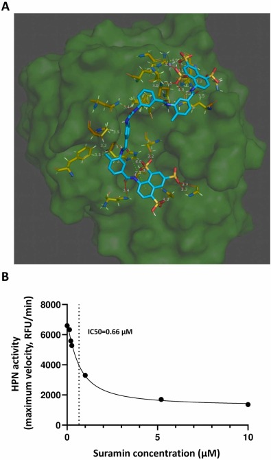
Fig. 1 Hepsin inhibition by suramin. A) Suramin blocks access to the hepsin catalytic site. Hepsin is shown as a green background. The suramin backbone is shown in light blue. Hepsin residues interacting with suramin are shown in yellow. Hydrophobic interactions are shown as gray dashed lines, hydrogen bonds as orange and red dashed lines, salt bonds as yellow dashed lines, and the pi-stacking interaction as a green dashed line. The numbers next to each interaction correspond to the distance in Angstroms of the bond. B) In the fluorogenic assay, the proteolytic activity of hepsin decreases with increasing suramin concentration. IC50: suramin concentration required to reduce the proteolytic activity of HPN by 50 %; RFU/min: relative fluorescence units/minute.
Image
Figure Caption
Acknowledgments
This image is the copyrighted work of the attributed author or publisher, and
ZFIN has permission only to display this image to its users.
Additional permissions should be obtained from the applicable author or publisher of the image.
Full text @ Biomed. Pharmacother.

