
|
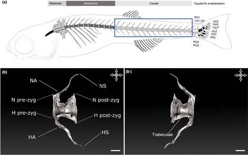
Fig. 1 Schematic of an adult zebrafish highlighting the region of interest (a; modified from Bensimon-Brito, Cardeira, et al., 2012) and SRXTM reconstructions of a normal vertebral body (b and b′). (b) Left side, (b′) right side. Scale bars = 100 μm. PU1 + U1, compound centrum preural 1 and ural 1; PU 2–3, preural 2–3; U2, ural 2; NA, neural arch; NS, neural spine; HA, haemal arch; HS, haemal spine; N and H pre-zyg, neural and haemal pre-zygapophysis; N and H post-zyg, neural and haemal post-zygapophysis. Please note that slight shape alterations not indicated by codes are related to the critical-point-drying procedure of previously whole-mount stained samples. The four-headed arrows indicate sample orientations.