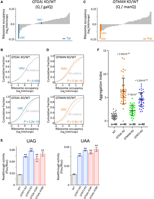
Fig. 5 Q-glycosylations regulate codon-specific translation and proteostasis (A and C) Fold-changes in ribosome occupancy at the A-site codon of QTGAL KO#1 (A) and QTMAN KO#1 (C), compared with WT HEK293T cells. Tyr and Asp codons are colored blue (A) and orange (C), respectively. Other codons are colored gray. (B and D) Cumulative plot of fold-changes in A-site ribosome occupancy of codons for Tyr (B, blue), Asp (D, orange), and others (gray) in QTGAL KO#1 (B) and QTMAN KO#1 (D), compared with WT HEK293T cells. p values were calculated by Mann-Whitney U test. (E) Dual luciferase reporter assay to examine readthrough of UAG (left) and UAA (right) codons in the respective KO cells. Each data point plotted on the bar graph shows mean ± SD (n = 3). ∗∗p < 0.01 by Student’s t test. (F) Measurement of cellular proteostasis in QTGAL KO and QTMAN KO using the EGFP-tagged firefly luciferase (Fluc) variant (R188Q). Aggregation index was calculated from the reporter protein aggregates divided by the area of EGFP fluorescence in microscopy images. Horizontal lines in the scattered dot plot represent mean ± SD. p values were calculated by Mann-Whitney U test. Datasets for WT and QTRT2 KO were referred to our previous study.26 See also Figure S3.
Reprinted from Cell, 186(25), Zhao, X., Ma, D., Ishiguro, K., Saito, H., Akichika, S., Matsuzawa, I., Mito, M., Irie, T., Ishibashi, K., Wakabayashi, K., Sakaguchi, Y., Yokoyama, T., Mishima, Y., Shirouzu, M., Iwasaki, S., Suzuki, T., Suzuki, T., Glycosylated queuosines in tRNAs optimize translational rate and post-embryonic growth, 5517-5535.e24, Copyright (2023) with permission from Elsevier. Full text @ Cell

