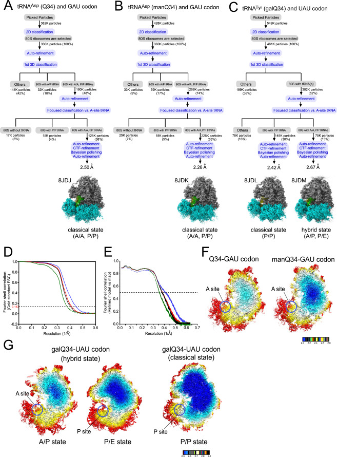
Fig. S4 Image processing procedures for cryo-EM of human 80S ribosomes, related to Figure 6 Image processing of human 80S ribosomes complexed with Q-tRNAs at both A- and P-sites. (A–C) (A) Human tRNAAsp bearing Q34 and the GAU codon at the A- and P-sites (PDB: 8JDJ , EMDB: EMD-36178), (B) Human tRNAAsp bearing manQ34 and the GAU codon at the A- and P-sites (PDB: 8JDK, EMDB: EMD-36179), (C) human tRNATyr bearing galQ34 and the UAU codon at the P-site in the classical state (PDB: 8JDL, EMDB: EMD-36180), and at the A- and P-sites in the hybrid state (PDB: 8JDM, EMDB : EMD-36181). The subgroup “others” includes A-site vacant 80S and low-resolution particles. (D and E) Gold standard Fourier shell correlation (FSC) curves (D) and refined model versus map FSC curves (E) of cryo-EM models. Black, tRNAAsp (Q34) with the GAU codon (PDB: 8JDJ , EMDB: EMD-36178), blue, tRNAAsp (manQ34) with the GAU codon (PDB: 8JDK, EMDB: EMD-36179), Red, tRNATyr (galQ34) with the UAU codon in the classical state (PDB: 8JDL, EMDB: EMD-36180), Green, tRNATyr (galQ34) with the UAU codon in the hybrid state (PDB: 8JDM, EMDB : EMD-36181). (F) Color-coded local resolution distribution of cryo-EM maps of human 80S ribosome complexed with tRNAAsp bearing Q34 (left) and manQ34 (right). Red circles indicate the A-sites. (G) Color-coded local resolution distribution of cryo-EM maps of human 80S ribosome complexed with tRNATyr bearing galQ34 in the hybrid state (left and middle) and in the classical state (right). Red circles indicate the P- and A-sites.
Reprinted from Cell, 186(25), Zhao, X., Ma, D., Ishiguro, K., Saito, H., Akichika, S., Matsuzawa, I., Mito, M., Irie, T., Ishibashi, K., Wakabayashi, K., Sakaguchi, Y., Yokoyama, T., Mishima, Y., Shirouzu, M., Iwasaki, S., Suzuki, T., Suzuki, T., Glycosylated queuosines in tRNAs optimize translational rate and post-embryonic growth, 5517-5535.e24, Copyright (2023) with permission from Elsevier. Full text @ Cell

