
|
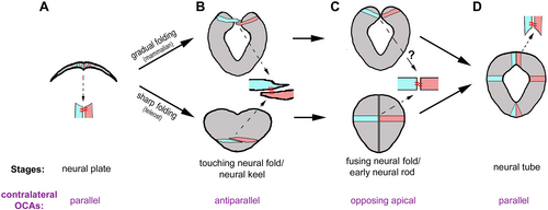
Fig. 3 The conservation of cellular configurations and the orientational cell adhesions (OCAs) between contralateral cells in neural tube closure. (A) At the neural plate stage, all cells are oriented with their apical ends facing dorsally. This configuration implies that only parallel OCAs exist between neighbouring contralateral cells that flank the midline. This cellular configuration and OCAs are similar among all vertebrates. (B) Gradual folding and sharp folding change the neural plate to the neural fold in mammals (top) and the neural keel in teleosts (bottom). The two folding modes imply that the conserved antiparallel intercellular configuration exists either narrowly in the dorsal region of the neural fold or broadly in the neural keel. (C) The antiparallel intercellular relationship recedes and gives rise to an opposing apical intercellular relationship, with contralateral cells coalesced at the apices through opposing apical OCAs. Opposing apical OCAs require N-cadherin in zebrafish. Whether opposing apical OCAs exist in mammals is yet to be determined. (D) At the neural tube stage, neighbouring contralateral cells have coalesced at the dorsal roof and ventral floor by only parallel OCAs.