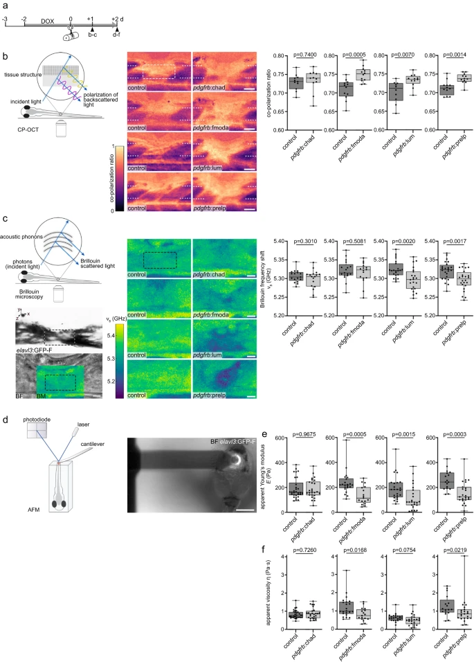
Fig. 7 SLRPs modulate the structural and mechanical properties of the lesion environment a Timeline for experimental treatments shown in b–f. b Targeting Fmoda, Lum, or Prelp to the injury ECM in pdgfrb:SLRP transgenic zebrafish increases the co-polarization ratio in the spinal lesion site, as determined by cross-polarized optical coherence tomography (CP-OCT). Images shown are average intensity projections of the lesion site (lateral view; rostral is left). The dashed lines indicate the location of the severed spinal cord, the dashed rectangle indicates the region of quantification. Two-tailed Student’s t-test. ncontrol = 11, nChad = 11; ncontro = 12, nFmoda = 12; ncontro = 10, nLum = 12; ncontrol = 12, nPrelp = 12 animals. c Targeting Lum or Prelp to the injury ECM in pdgfrb:SLRP transgenic zebrafish decreases the Brillouin frequency shift ( ) in the spinal lesion site, as determined by Brillouin microscopy. Images shown are sagittal optical sections (brightfield intensity, confocal fluorescence, or Brillouin frequency shift map) through the center of the lesion site of an elavl3:GFP-F transgenic zebrafish (lateral view; rostral is left). The dashed rectangle indicates the region of quantification. Two-tailed Student’s t-test. ncontrol = 17, nChad = 17; ncontrol = 17, nFmoda = 16; ncontrol = 23, nLum = 22; ncontrol = 24, nPrelp = 24 animals over three (Chad, Fmoda) or four (Lum, Prelp) independent experiments. d–f Targeting Fmoda, Lum, or Prelp to the injury ECM in pdgfrb:SLRP transgenic zebrafish decreases the apparent Young’s modulus (E) (e; Fmoda, Lum, Prelp) and apparent viscosity (η) (f; Fmoda, Prelp) in the spinal lesion site, as determined by atomic force microscopy-based nanoindentation measurements (AFM). Recorded force-indentation curves were fitted to the Kelvin─Voigt─Maxwell model. Two-tailed Mann-Whitney test. ncontrol = 30, nChad = 26; ncontrol = 22, nFmoda = 17; ncontrol = 24, nLum = 23; ncontrol = 17, nPrelp = 23 animals over three (Chad, Fmoda, Prelp) or four (Lum) independent experiments. a–f Each data point represents one animal. Box plots show the median, first, and third quartile. Whiskers indicate the minimum and maximum values. Scale bars: 500 µm (d), 50 µm (b), 10 µm (c). BF brightfield intensity, BM Brillouin frequency shift map, d days, dpl days post-lesion, DOX doxycycline. Source data are provided as a Source Data file.
Image
Figure Caption
Acknowledgments
This image is the copyrighted work of the attributed author or publisher, and
ZFIN has permission only to display this image to its users.
Additional permissions should be obtained from the applicable author or publisher of the image.
Full text @ Nat. Commun.

