
Image
|
Figure Caption
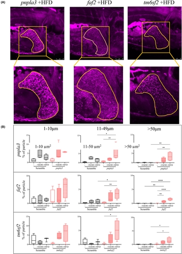
Fig. 5 Nile Red staining showed larger size lipid droplets in 2% ethanol- or HFD-treated crispants. (A) Representative images show the liver area selected to calculate the lipid droplets size count in pnpla3, faf2 and tm6sf2 crispants treated with HFD, stained with Nile Red (magenta) and imaged using confocal microscopy. (B) Percentage of number of lipid droplet size (μm2), 1–10, 10–50 and greater 50 was measured using Fiji ImageJ software in scramble control and pnpla3, faf2 and tm6sf2 larvae on 5 dpf following 48 h of exposure to 2% EtOH and HFD.
Acknowledgments
This image is the copyrighted work of the attributed author or publisher, and
ZFIN has permission only to display this image to its users.
Additional permissions should be obtained from the applicable author or publisher of the image.
Full text @ Liver Int.