
IMAGE
Figure 1
- ID
- ZDB-IMAGE-240112-42
- Genes
- Publication
- Raman et al., 2023 - A Zebrafish Mutant in the Extracellular Matrix Protein Gene efemp1 as a Model for Spinal Osteoarthritis
- All Figures
- Figures for Raman et al., 2023
Image
|
Figure Caption
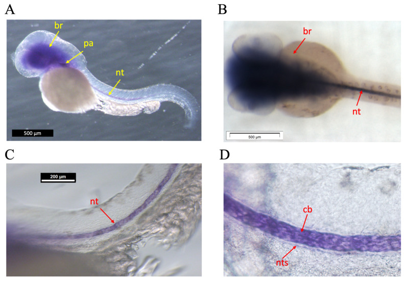
Figure 1
Whole mount in situ hybridization on 48 hpf zebrafish embryos. (
Figure Data
Acknowledgments
This image is the copyrighted work of the attributed author or publisher, and
ZFIN has permission only to display this image to its users.
Additional permissions should be obtained from the applicable author or publisher of the image.
Full text @ Animals (Basel)