
Image
|
Figure Caption
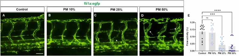
Fig. 4 Vasculature malformation and defect in blood flow induced by PM exposure. (A–D) Representative images of the blood vasculature of Tg(fli1a:egfp) zebrafish embryos at 3 dpf. White arrows indicate the intersegmental vessels with abnormal branches. Scale bar, 50 μM. (E) Flow rate of blood cells in PM-exposed zebrafish embryos. Kruskal–Wallis test (n = 16 per group; ***p < 0.001 and ****p < 0.0001; n.s., not significant.).
Acknowledgments
This image is the copyrighted work of the attributed author or publisher, and
ZFIN has permission only to display this image to its users.
Additional permissions should be obtained from the applicable author or publisher of the image.
Full text @ Ecotoxicol. Environ. Saf.