
|
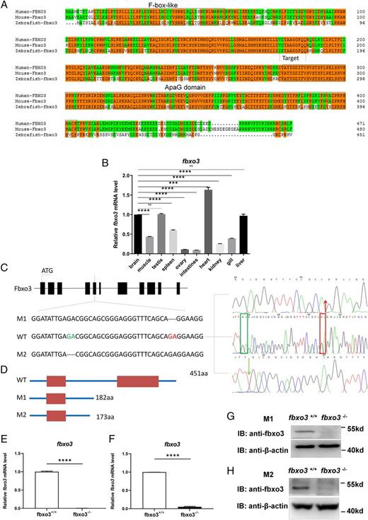
Fig. 4 Generation of fbxo3-null zebrafish via CRISPR/Cas9 technology. (A) Amino acid sequence alignment of human, mouse, and zebrafish fbxo3. Human FBXO3 (ENSG00000110429), mouse Fbxo3 (ENSMUSG00000027180), zebrafish fbxo3 (ENSDARG00000029242); the same amino acids are highlighted with orange, the F-box–like domain and ApaG domain are circled with blue boxes. (B) Expression pattern of fbxo3 in different tissues of adult zebrafish (3 mpf) as revealed by qRT-PCR assays. (C) Scheme of the genomic structure of zebrafish fbxo3. The sequence information of fbxo3 zebrafish mutant: fbxo3ihb0703/ihb0703 (M1), 2 bp nt (GA) were deleted from exon 5 of fbxo3, resulting in a reading frame shift and generating truncated proteins with 182 aa; fbxo3ihb1224/ihb1224 (M2), 2 bp nt (GA) were deleted from exon 5 of fbxo3, resulting in a reading frame shift and generating truncated proteins with 173 aa. (D) Scheme of the putative peptides of the WT transcript and the two mutated fbxo3 transcripts. (E) Expression of fbxo3 mRNA in larvae from the WT (fbxo3+/+) and the fbxo3-null (fbxo3ihb0703/ihb0703) zebrafish (4 dpf; n = 50, respectively). (F) Expression of fbxo3 mRNA in larvae from the WT (fbxo3+/+) and the fbxo3-null (fbxo3ihb1224/ihb1224) zebrafish (4 dpf; n = 50, respectively). (G) Expression of fbxo3 protein in brain from the WT (fbxo3+/+) and the fbxo3-null (fbxo3ihb0703/ihb0703) zebrafish (2 mpf). (H) Expression of fbxo3 protein in brain from the WT (fbxo3+/+) and the fbxo3-null (fbxo3ihb1224/ihb1224) zebrafish (2 mpf). Data are presented as means ± SEM of three biological replicates. ***p < 0.001, ****p < 0.0001. ns, no significance.