
Fig. 3
- ID
- ZDB-IMAGE-231228-142
- Publication
- Torraca et al., 2023 - Zebrafish null mutants of Sept6 and Sept15 are viable but susceptible to Shigella infection
- All Figures
- Figures for Torraca et al., 2023
|
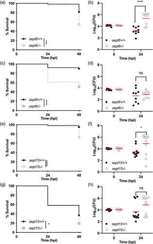
Fig. 3 Zebrafish null mutants of Sept6 and Sept15 are more susceptible to Shigella infection. (a–d) Survival curves (a,c) and Log10-transformed CFU counts (b,d) of 3 dpf sept6+/+ and sept6−/− larvae (wild-type in black; mutant in white) injected in the hindbrain ventricle (HBV) with S. flexneri GFP+ at a dose of 10,000 CFUs (a,b) or a dose of 5,000 CFUs (c,d). Next, larvae were incubated at either 28.5°C (a,b) or 32.5°C (c,d), for up to 48 h. Experiments are cumulative of three biological replicates. Sample size: For survival analysis, a total of 65 (wild-type) and 79 (mutant) and 90 (wild-type) and 80 (mutant) larvae was analysed at 28.5°C and 32.5°C, respectively. For CFU analysis, a total of 9 larvae was analysed per experimental group. Only living larvae were used for CFU enumeration. Statistics: Log-rank (Mantel-Cox) test (a,c); unpaired t test on Log10-transformed values (b,d). ****p < 0.0001; ns p > 0.05, respectively. (e,h) Survival curves (e,g) and Log10-transformed CFU counts (f,h) of 3 dpf sept15+/+ and sept15−/− larvae (wild-type in black; mutant in white) injected in the HBV with S. flexneri GFP+ at a dose of 10,000 CFUs. Next, larvae were incubated at either 28.5°C (e,f) or 32.5°C (g,h) for up to 48 h. Experiments are cumulative of four biological replicates. Sample size: For survival analysis, a total of 99 (wild-type) and 104 (mutant) and 103 (wild-type) and 113 (mutant) larvae was analysed at 28.5°C and 32.5°C, respectively. For CFU analysis, a total of 12 larvae was analysed per experimental group. Only living larvae were used for CFU enumeration. Statistics: Log-rank (Mantel–Cox) test (e, g); unpaired t test on Log10-transformed values (f,h). *p < 0.05; ****p < 0.0001; ns p > 0.05, respectively