
|
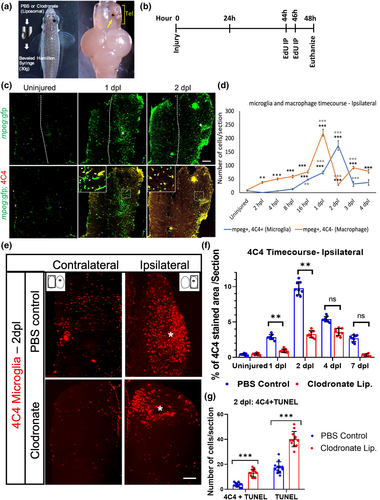
Fig. 1 Liposomal clodronate suppresses activated microglia after telencephalic injury. (a) Description of the lesioning method and injury location in the right telencephalon (asterisk, left panel), and an example of a dissected zebrafish brain showing the injury (yellow arrow, right panel). Tel, telencephalon. (b) Timeline of lesioning, EdU labeling and euthanasia for the 2-day time point. (c) Microglial response after injury at 1 and 2 days post lesion (dpl) as measured by tg(mpeg:GFP) reporter expression alone (green, top panels) or after double-labeling with 4C4 antibody to identify activated microglia (red, bottom panels). The midline is indicated by the dotted lines and injured regions by asterisks in the top panels. (d) Quantification of mpeg:GFP/4C4 double-positive microglia (blue line) or mpeg:GFP+/4C4− macrophages (red line) from 2 hours post lesioning (hpl) to 4 dpl. **p < .001; ***p < .0001 by repeated measures ANOVA followed by Tukey's post-hoc test. Comparisons for each curve were made between uninjured animals (the first points on the curve) and lesioned fish at each timepoint with statistical significance denoted by black asterisks, and between lesioned fish at a given timepoint versus the previous timepoint, with gray asterisks denoting significant differences. (e) 4C4 immunolabeling at 2 dpl after injection of control (top panels) or clodronate (bottom panels) liposomes. * Indicates the site of injury. (f) Quantification of 4C4+ microglia in the ipsilesional telencephalon at 1, 2, 4, and 7 dpl in control and clodronate groups. n = 12, **p < .001 by repeated measure ANOVA with Tukey's post-hoc test. (g) Quantification of 4C4 and TUNEL labeling at 2 dpl in control and clodronate groups. ***p < .0001 by t-test. Scale bar = 10 μm for (c and e); error bars indicate SEM for all graphs.