
|
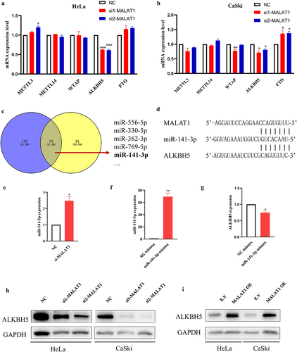
Fig. 3 Effect of MATAL1on ALKBH5 expression in HeLa and CaSki cells. (a and b) NC and two siRNA (si1 and si2) for silencing MALAT1 were transfected into HeLa and CaSki cells. The expression of the m6A-associated genes (METTL3, METTL14, WTAP, ALKBH5, and FTO) was determined by RT-qPCR. (c) The candidate miRNAs, which might bind to MALAT1 or ALKBH5, were predicted by StarBase. (d) Sequence of miR-141-3p was matched with MALAT1 and ALKBH5 3’UTR. (e) The expression of miR-141-3p was analyzed after knockdown of MALAT1 in HeLa cells. (f) The expression of miR-141-3p was analyzed after transient transfection of miR-141-3p mimics in HeLa cells using RT-qPCR. (g) The mRNA expression level of ALKBH5 was analyzed by RT-qPCR following transfection of miR-141-3p mimics in HeLa cells. (h) The cell lysates were subjected to western blot analysis for ALKBH5 expression after silencing MALAT1 using GAPDH as loading control. (i) The MALAT1 was overexpressed followed by analysis of ALKBH5 protein expression in HeLa and CaSki cells. NC, negative control.