
|
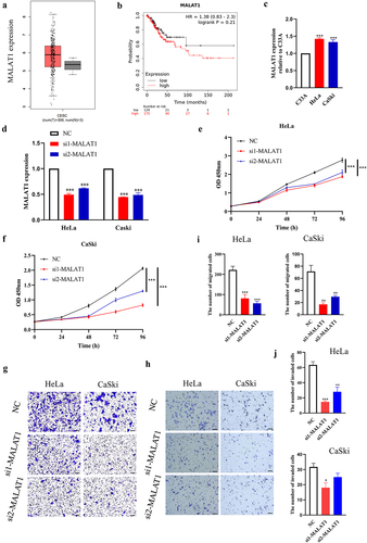
Fig. 1 Expression and effect of MALAT1 on cervical cancer cells in vitro. (a) The expression of MALAT1 in tumor samples was analyzed. (b) The overall survival rate of patients with high and low MALAT1 expression was analyzed. (c) The expression level of MALAT1 was determined by RT-qPCR. Fold changes are shown using C33A cells as control. (d) HeLa and CaSki cells were transfected with NC and two specific siRNAs against MALAT1 (si1 and si2), and the expression of MATAL1 was measured using RT-qPCR. Quantitative data are shown. (e and f) Cells were treated the same as described in (d), and cell growth assay was performed at different time points, as indicated. (g-j) Cell migration and invasion assay were performed after transfection of siRNAs for MATAL1 in Hela and CaSki cells, respectively. The representative fields were photographed (G : migration, H: invasion) (scale bar: 100 μm), and the numbers of migrated and invaded cells were calculated. Three independent experiments were performed. NC, negative control.