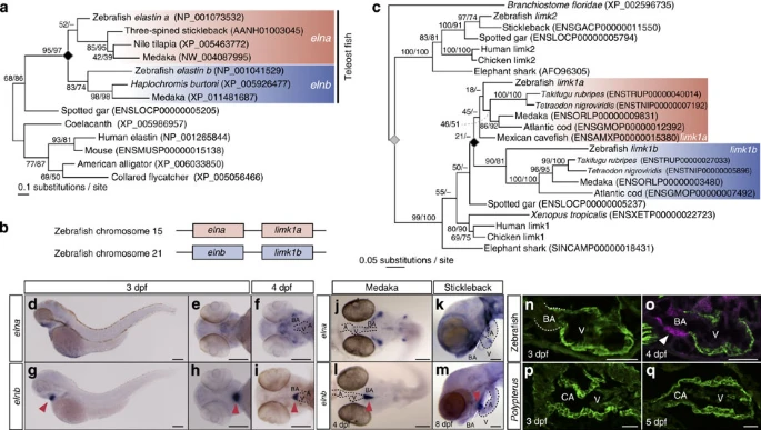
Fig. 2 Molecular phylogeny and expression patterns of elna and elnb in teleost.(a) Molecular phylogenetic tree of elastin genes (elna/elnb) inferred using 83 amino-acid sites (shape parameter of the gamma distribution alpha=0.67). The gene duplication between elna and elnb is indicated with the black diamond. (b) Syntenic relationship of elna/elnb and limk1a/1b. (c) Molecular phylogenetic tree of limk1 genes inferred using 303 amino-acid sites (alpha=1.03). The gene duplication between limk1a and limk1b is indicated with the black diamond, while a more ancient gene duplication is shown with the grey diamond. The limk1a-1b duplication is shown to have occurred early in the teleost fish lineage, coinciding with 3R WGD. (d–i) Expression patterns of elna and elnb in zebrafish embryos at 3 dpf (d,e,g,h) and 4 dpf (f,i). Arrowheads indicate signals in the BA. Scale bars, 200 μm. (j–m) Expression patterns of elna and elnb in medaka (j,l) and stickleback (k,m) embryos. Note that elnb is expressed only in the BA in both medaka and stickleback embryos. Scale bars, 200 μm. (n–q) Double immunohistological staining of developing OFTs in zebrafish (n,o) and Polypterus (p,q) embryos against α-sarcomeric actinin (cardiac muscle, green) and myosin light-chain kinase (smooth muscle, magenta). Smooth muscle cells appear in the zebrafish BA at 4 dpf, but could not be detected in the Polypterus CA. A, atrium. Scale bars, 50 μm.
Image
Figure Caption
Figure Data
Acknowledgments
This image is the copyrighted work of the attributed author or publisher, and
ZFIN has permission only to display this image to its users.
Additional permissions should be obtained from the applicable author or publisher of the image.
Full text @ Nat. Commun.

