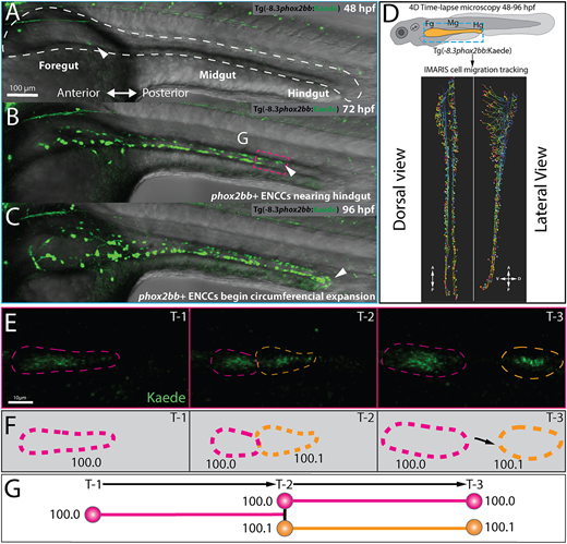
Fig. 1 Single-cell generation tracing of ENCCs during early ENS development in toto. (A-C) Still images from time-lapse reveal phox2bb+ ENCCs emerging into foregut intestine at 48 hpf (A, white arrow), leading ENCCs reaching the hindgut at 72 hpf (B, white arrow) and the circumferential expansion of ENCCs at 96 hpf (C). Dashed white line in A shows an outline of the gut tube region of interest. (D) Schematic (top) illustrates region of interests for time-lapse microscopy of ENCC migration in intestine and lateral and dorsal view snapshots (bottom) of single-cell migration tracking in Imaris. (E) Expanded images of cell migration and division at the ENCC wavefront (boxed area, B). Time-points 1-3 (T1-3) depict representative time-points at which cell divisions occur. (F) Cartoon corresponds to cell outlines in panel E (dashed lines) and with representative unique ID labels that denote cell generation number as a decimal. (G) Cartoon generation-tree corresponds to cell divisions seen in panels E and F and depict how unique IDs are applied to cells throughout the time-lapse. A, anterior; D, dorsal; P, posterior; V, ventral.
Image
Figure Caption
Figure Data
Acknowledgments
This image is the copyrighted work of the attributed author or publisher, and
ZFIN has permission only to display this image to its users.
Additional permissions should be obtained from the applicable author or publisher of the image.
Full text @ Development

