
|
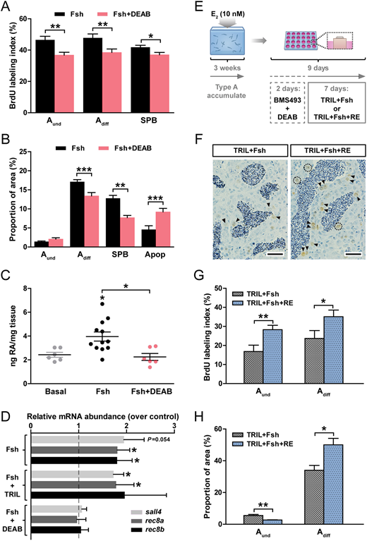
Fig. 7 RA signaling contributes to Fsh-stimulated spermatogonial differentiation. Determination of BrdU labeling indices (A) and of the frequency (B) of type A and B spermatogonia and apoptotic cells. Testicular explants were cultured for 4 days under Fsh-stimulated (100 ng/ml) conditions, in the absence or presence of 10 µM DEAB. (C) Quantification of RA production by testis tissue cultured for 4 days in response to basal medium, 100 ng/ml Fsh alone or with 10 µM DEAB. (D) Expression of RA target genes in testicular explants cultured for 4 days under Fsh-stimulated (100 ng/ml) conditions, in the absence or presence of 25 µg/ml TRIL or of 10 µM DEAB. (E-H) Retinoid signaling directly stimulates spermatogonial proliferation during spermatogenetic recovery. (E) Testis tissue was collected from adult males exposed to 10 nM estradiol (E2) in vivo for 21 days and then cultured for 2 days in the presence of BMS493 (10 µM) and DEAB (10 µM) inhibitors, followed by another 7 days incubation with different treatments (25 µg/ml TRIL plus 100 ng/ml Fsh) in the absence or presence of 10 µM RE. (F-H) Detection of BrdU (F) and quantification of BrdU labeling indices (G) and the areas occupied by type A spermatogonia (H). Data are expressed as mean±s.e.m. (n=5-12; *P<0.05, **P<0.01, ***P<0.001). In D, results are shown as relative to the basal control condition, which is set at 1 (dashed line). Scale bars: 25 µm in F. BrdU-positive Aund cells are indicated by dashed black lines, Adiff by arrowheads.