
Fig. 4
- ID
- ZDB-IMAGE-231215-4
- Publication
- Tu et al., 2022 - Dhx38 regulates the maintenance and differentiation of erythro-myeloid progenitors and hematopoietic stem cells by alternative splicing
- All Figures
- Figures for Tu et al., 2022
|
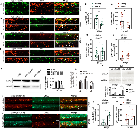
Fig. 4 Dhx38 deficiency induces abnormal mitosis and apoptosis of EMPs and HSPCs in zebrafish. (A) EdU assay in Tg(cmyb:eGFP) indicates an increase of cmyb+ cells in the S phase of dhx38−/− embryos at 36 hpf. White arrows indicate the colocalization of cmyb and EdU. Sibling, n=8; dhx38−/−, n=10; performed with three replicates; **P=0.001. (B) EdU assay in Tg(gata1:dsred) indicates an increase in the number of gata1+ cells in the S phase in dhx38−/− embryos at 36 hpf. White arrowheads indicate the colocalization of gata1 and EdU. Sibling, n=17; dhx38−/−, n=12; performed with three replicates; *P=0.029. (C,D) Quantification of cmyb+ EdU+ cells and gata+ EdU+ cells from A,B, respectively. (E) Double immunostaining of Tg(cmyb:eGFP) and pH3 shows that the number of cmyb+ cells in the M phase is elevated in the dhx38 mutants at 36 hpf. White arrowheads indicate the colocalization of cmyb and pH3. sibling, n=13; dhx38−/−, n=12; performed with three replicates; *P=0.028. (F) Double immunostaining of Tg(gata1:dsred) and pH3 shows that the number of gata1+ cells in the M phase is also elevated in dhx38 mutants at 36 hpf. White arrowheads indicate the colocalization of gata1 and pH3. Sibling, n=17; dhx38−/−, n=24; performed with three replicates; **P=0.0025. (G,H) Quantification of cmyb+ pH3+ cells and gata+ pH3+ cells from E,F, respectively. (I) The siDHX38-641 siRNA is at the c.641 position of the human DHX38 gene, and siDHX38-328 is at c.328. The silencing efficiency of these two siRNAs was confirmed by western blotting (left). Western blotting for DHX38 shows the efficiency of DHX38 knockdown by si-DHX38-641, but not by si-DHX38-325 (middle). Flow cytometry analysis of the cell cycle (right) after treatment with si-DHX38-641, showing a decrease of cells in the S phase and an increase of cells in the M phase. Performed with six replicates; n.s., P=0.18; **P=0.001; ***P=0.0001. (J) The protein levels of γH2AX in siblings and dhx38−/− zebrafish at 36 and 56 hpf were detected by western blotting. GAPDH was used to normalize protein loading. n=3; for 36 hpf, *P=0.01; for 48 hpf *P=0.015. (K,L) TUNEL assay in Tg(cmyb:eGFP) (bottom) and Tg(gata1:dsred) (top) shows that apoptotic cmyb+ and gata1+ cells are increased in the dhx38 mutants at 56 hpf. The double-positive fluorescence demonstrates that cmyb+ and gata1+ cells underwent apoptosis. For Tg(gata1:dsred): sibling, n=12; dhx38−/−, n=13; performed with three replicates; *P=0.019. For Tg(cmyb:eGFP): sibling, n=9; dhx38−/−, n=9; performed with three replicates; ****P=0.00006. (M,N) Quantification of double-positive fluorescent cell number from K,L. Data show the mean±s.d. Significance was determined using a two-tailed, unpaired Student's t-test. n.s., not significant; *P<0.05; **P<0.01; ***P<0.001; ****P<0.0001. All scale bars: 50 μm.