
Fig. 3
- ID
- ZDB-IMAGE-231215-3
- Publication
- Tu et al., 2022 - Dhx38 regulates the maintenance and differentiation of erythro-myeloid progenitors and hematopoietic stem cells by alternative splicing
- All Figures
- Figures for Tu et al., 2022
|
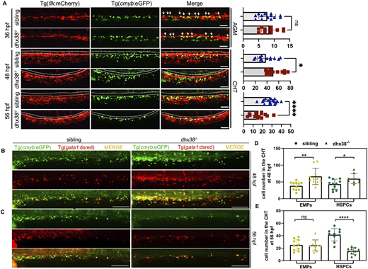
Fig. 3 Loss of dhx38 impairs definitive hematopoiesis. (A) In vivo imaging of hematopoietic progenitor cells in Tg(cmyb:eGFP;flk:mCherry) at 36 hpf, 48 hpf and 56 hpf. The double-positive fluorescence (white arrows) in the VDA region shows that hemogenic endothelium emerges normally in the dhx38 mutants. However, the number of cmyb+ cells in the dhx38 mutants are increased at 48 hpf, but decreased at 56 hpf. White dotted lines represent the caudal artery. For 36 hpf: sibling, n=13; dhx38−/−, n=13; P=0.51, not significant. For 48 hpf: sibling, n=14; dhx38−/−, n=19; *P=0.016. For 56 hpf: sibling, n=19; dhx38−/−, n=15; ****P=0.000023. Quantification of cells from 36 hpf, 48 hpf, and 56 hpf is shown on the right. (B) Immunostaining of Tg(cmyb:eGFP;gata1:dsred) fish at 48 hpf shows an increase in the number of cmyb+/gata1+ cells (EMPs) and cmyb+/gata1− cells (HSPCs) in the CHT of dhx38 mutants. (C) Immunostaining of Tg(cmyb:eGFP;gata1:dsred) fish at 56 hpf demonstrates a decreased number of cmyb+/gata1+ EMPs and cmyb+/gata1− HSPCs in the CHT of dhx38 mutants. (D) Quantification of cmyb+/gata1+ and cmyb+/gata1− cell numbers from B. Sibling, n=11; dhx38−/−, n=11; EMPs, **P=0.0026; HSPCs, *P=0.031. (E) Quantification of cmyb+/gata1+ and cmyb+/gata1− cell number from C. Sibling, n=10; dhx38−/−, n=10; EMPs, P=0.86; HSPCs, ****P= 0.0000009. Data show the mean±s.d. Significance was determined using a two-tailed, unpaired Student's t-test. n.s., not significant; *P<0.05; **P<0.01; ****P<0.0001. All scale bars: 200 μm.