
Fig. 2
- ID
- ZDB-IMAGE-231215-2
- Publication
- Tu et al., 2022 - Dhx38 regulates the maintenance and differentiation of erythro-myeloid progenitors and hematopoietic stem cells by alternative splicing
- All Figures
- Figures for Tu et al., 2022
|
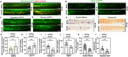
Fig. 2 Maturation of EMPs is perturbed in dhx38 mutants. (A) In vivo imaging of gata1+/cmyb+ cells (EMPs) in the PBI region of Tg(cmyb:eGFP;gata1:dsred) fish. At 36 hpf and 48 hpf, the numbers of EMPs (indicated in yellow) in the dhx38−/− zebrafish are higher compared with wild-type siblings. Sibling, n=16; dhx38−/−, n=16; performed with three replicates; **P=0.002. (B) In vivo imaging of Tg(pu.1:eGFP) fish shows that promyelocytes are increased in the PBI region of dhx38 mutants at 36 hpf and 48 hpf. Sibling, n=12; dhx38−/−, n=10; performed with three replicates; ***P=0.0006. (C) In vivo imaging of macrophages in Tg(mpeg1:eGFP) also shows a decreased branched cell number in the dhx38 mutant at 48 hpf. Sibling, n=11; dhx38−/−, n=11; performed with three replicates; ****P=0.00003. (D) In vivo imaging of granulocytes in Tg(mpx:eGFP) displays a decreased number in the dhx38 mutant at 48 hpf. Sibling, n=12; dhx38−/−, n=11; performed with three replicates; ***P=0.00013. (E) Sudan Black staining in dhx38−/− zebrafish shows that mature granulocytes are decreased at 48 hpf. Black arrows indicate granulocytes. Sibling, n=34; dhx38−/−, n=23; performed with three replicates; ****P=0.000001. (F) Neutral Red staining shows significantly decreased numbers of functional macrophages. Black arrows indicate macrophages. Sibling, n=14; dhx38−/−, n=11; performed with three replicates; ***P=0.00014. (G-L) Quantification of cells from A-G. Significance was determined using a two-tailed, unpaired Student's t-test. **P<0.01; ***P<0.001; ****P<0.0001. All scale bars: 50 μm.