
Fig. 5
- ID
- ZDB-IMAGE-231215-154
- Publication
- Touret et al., 2022 - A dual involvement of Protocadherin-18a in stromal cell development guides the formation of a functional hematopoietic niche
- All Figures
- Figures for Touret et al., 2022
|
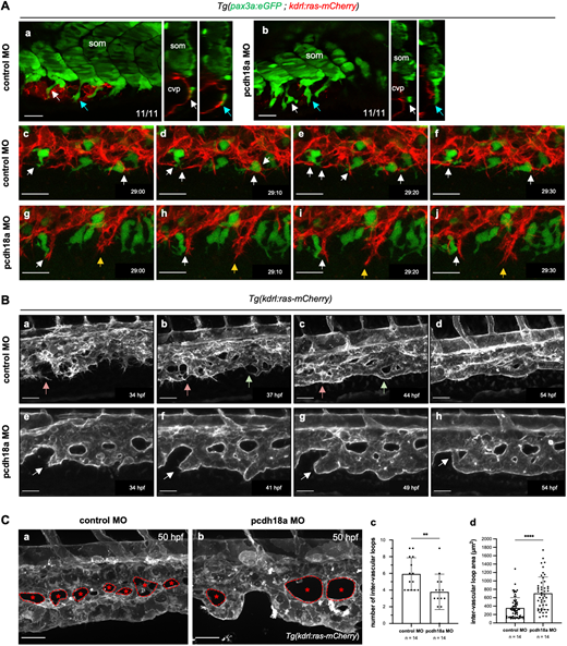
Fig. 5 Pcdh18a truncation impacts on venous plexus morphogenesis. (Aa,Ab) Confocal single-slice images and orthogonal (transverse) views of control (Aa) and pcdh18a (Ab) MO-injected Tg(pax3a:eGFP; kdrl:ras-mCherry) embryos at 26 hpf. White and blue arrows point to the same cells (SCPs) shown in transverse view on the right; n=11 control and 11 pcdh18a MO-injected embryos, from four independent experiments. cvp, caudal vein plexus; som, somite. (Ac-Aj) Time-lapse confocal imaging of the forming CVP of control (Ac-Af) and pcdh18a-ΔCP106 morphant (Ag-Aj) embryos (see also Movie 5). Images were extracted at 10 min intervals from 29 hpf to 29.5 hpf. Dynamic interactions between stromal and endothelial cells (white arrows) were frequent in control embryos whereas in pcdh18a morphants endothelial cells migrated ventralwards apparently regardless of stromal cells (yellow arrows). Scale bars: 20 µm. (Ba-Bh) Time-lapse confocal images from control (Ba-Bd) and pcdh18a (Be-Bh) MO-injected Tg(kdrl:ras-mCherry) embryos at later stages (34-54 hpf). See also Movie 6. In the control, red and green arrows follow the formation of new vascular segments. In the morphant, white arrows point to the abnormal tubulogenesis of the definitive caudal vein. Scale bars: 30 µm. (C) Morphological differences in the IVLs. (Ca,Cb) Confocal spinning-disk acquisitions of live transgenic Tg(kdrl:ras-mCherry) control or pcdh18a MO-injected embryos at 50 hpf. IVLs are indicated by asterisks and the border of each IVL is delineated by a dashed line. Scale bars: 30 µm. (Cc,Cd) Number (Cc) and surface in µm2 (Cd) of IVLs over a five-somite width in controls and morphants; n=14 embryos per condition, from two independent experiments (mean±s.d.; **P=0.0066; ****P<0.0001; Mann–Whitney test).