
|
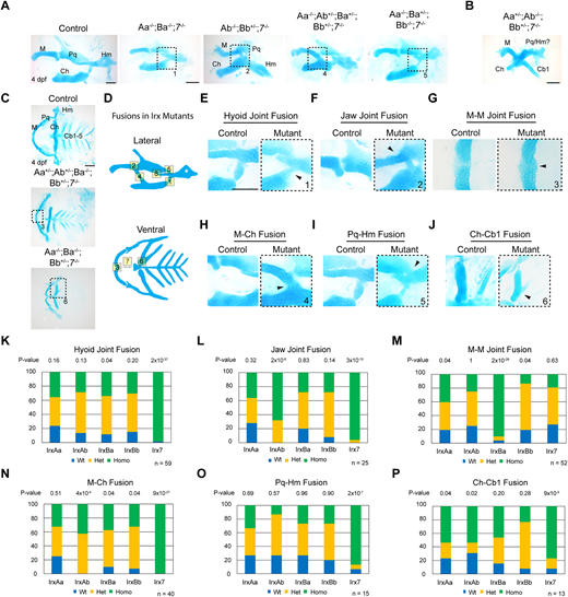
Fig. 6 Widespread facial cartilage fusions and joint loss in Irx mutants. (A,B) Dissected first and second arch-derived skeletons shown in lateral view and stained with Alcian Blue (cartilage) at 4 dpf. Distinct combinations of Irx mutations result in a range of cartilage fusions and joint loss (boxed regions) (A), and in severe cases fusions of multiple cartilages (B). (C) Dissected facial cartilages shown in ventral view, with dashed boxes showing cartilage fusions. (D) Schemes of the range of cartilage fusions and joint losses seen in Irx mutant combinations. Numbers correspond to the boxed regions in A and C; see Fig. S4 for fusions 7 and 8. (E-J) Magnifications of the cartilage fusions seen in A and C (boxed regions) versus similar regions from wild-type controls. (K-P) Summary of genotypes for the indicated phenotypes from a quintuple heterozygous in-cross. P-values report chi-square tests for deviation from the expected 1:2:1 wild type:heterozygous:homozygous Mendelian ratio. n denotes animals in which the indicated fusions were observed. Cb, ceratobranchial; Ch, ceratohyal; Hm, hyomandibular; M, Meckel's; Pq, palatoquadrate. Scale bars: 100 µm.