
IMAGE
Figure 3.
- ID
- ZDB-IMAGE-231212-44
- Publication
- Singh et al., 2023 - NMJ analyser: a novel method to quantify neuromuscular junction morphology in zebrafish
- All Figures
- Figures for Singh et al., 2023
Image
|
Figure Caption
Figure 3.
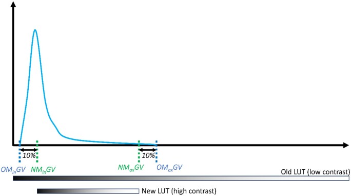
Distribution curve demonstrating histogram LUT compression. Increasing the minimum and decreasing the maximum grayscale values by 10% of the dynamic range (max. to min. grayscale value) of the image slice increases the contrast of the image for puncta detection. NMinGV: New Minimum Grayscale Value, NMaxGV: New Maximum Grayscale Value, OMinGV: Original Mininum Grayscale Value; OMaxGV: Original Maximum Grayscale Value.
Acknowledgments
This image is the copyrighted work of the attributed author or publisher, and
ZFIN has permission only to display this image to its users.
Additional permissions should be obtained from the applicable author or publisher of the image.
Full text @ Bioinformatics